Fig. 2
- ID
- ZDB-FIG-221023-14
- Publication
- Liu et al., 2022 - Lrpap1 deficiency leads to myopia through TGF-β-induced apoptosis in zebrafish
- Other Figures
- All Figure Page
- Back to All Figure Page
|
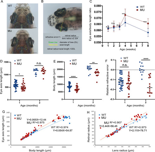
Longitudinal measurements of the eye dimensions using in vivo imaging. A Eye globes of wild-type and lrpap1 mutant zebrafish at three months. The scale bars refer to 500 μm. B The different ocular parameters measured and the method of calculation of the relative refractive error (RRE) used. C The eye axis to body length ratio was determined at different time points. The eye axis length (D) and body length (E) of one- and two-month-old wild-type and mutant zebrafish were also determined individually. F RRE measurements in one- and two-month-old mutant and wild-type zebrafish. G Correlation analysis of the eye axis length and body length measurements. H Correlation analysis of the retinal radius and lens radius measurements. WT, wild-type. MU, lrpap1 homozygous mutant. Statistical significance was determined using the Student’s t-test: n.s. = no significance; *p < 0.05; **p < 0.01; ***p < 0.001; ****p < 0.0001. For wild-type zebrafish, n = 16, 13, 12, 36, and 12 eyes one week, two weeks, three weeks, one month, and two months post-fertilization, respectively. For the lrpap1 mutant line, n = 12, 10, 13, 24, and 14 eyes one week, two weeks, three weeks, one month, and two months post-fertilization, respectively |
| Fish: | |
|---|---|
| Observed In: | |
| Stage Range: | Days 30-44 to Days 45-89 |

