Fig. 4
- ID
- ZDB-FIG-221001-32
- Publication
- Shorey et al., 2021 - Microtubule organization of vertebrate sensory neurons in vivo
- Other Figures
- All Figure Page
- Back to All Figure Page
|
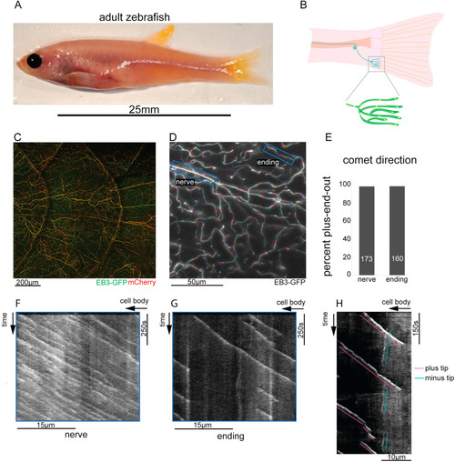
Fig. 4. Zebrafish DRG sensory endings have uniform plus-end-out microtubule polarity throughout their entire arbor. A. Brightfield overview of an 8-10 month-old adult zebrafish. B. Diagram of a single DRG sensory neuron innervating a posterior scale. C. Confocal z-stack of a scale innervated by DRG sensory neurons expressing mCherry and EB3-GFP D. False-colored time series z-stack image of the surface of a scale on an 8-10 month-old zebrafish expressing EB3-GFP with successive timepoints batched into colors progressing from the blue to red end of the spectrum, showing a series of rainbows with their red end oriented towards the end of the neurite, reflecting a plus end out microtubule polarity. E. Quantification of microtubule polarity from movies of EB3-GFP in DRG nerves and endings in 8–10 month-old zebrafish; number on column is the number of comets counted for that condition F.G. Representative kymographs of EB3-GFP in the nerve and sensory endings respectively. H. Kymograph generated from a movie of EB3-GFP in a sensory ending in which both growing minus ends (blue) and plus ends (pink) are visible. |
Reprinted from Developmental Biology, 478, Shorey, M., Rao, K., Stone, M.C., Mattie, F.J., Sagasti, A., Rolls, M.M., Microtubule organization of vertebrate sensory neurons in vivo, 1-12, Copyright (2021) with permission from Elsevier. Full text @ Dev. Biol.

