
|
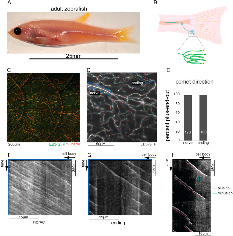
Fig. 4
Fig. 4. Zebrafish DRG sensory endings have uniform plus-end-out microtubule polarity throughout their entire arbor. A. Brightfield overview of an 8-10 month-old adult zebrafish. B. Diagram of a single DRG sensory neuron innervating a posterior scale. C. Confocal z-stack of a scale innervated by DRG sensory neurons expressing mCherry and EB3-GFP D. False-colored time series z-stack image of the surface of a scale on an 8-10 month-old zebrafish expressing EB3-GFP with successive timepoints batched into colors progressing from the blue to red end of the spectrum, showing a series of rainbows with their red end oriented towards the end of the neurite, reflecting a plus end out microtubule polarity. E. Quantification of microtubule polarity from movies of EB3-GFP in DRG nerves and endings in 8–10 month-old zebrafish; number on column is the number of comets counted for that condition F.G. Representative kymographs of EB3-GFP in the nerve and sensory endings respectively. H. Kymograph generated from a movie of EB3-GFP in a sensory ending in which both growing minus ends (blue) and plus ends (pink) are visible.
Reprinted from Developmental Biology, 478, Shorey, M., Rao, K., Stone, M.C., Mattie, F.J., Sagasti, A., Rolls, M.M., Microtubule organization of vertebrate sensory neurons in vivo, 1-12, Copyright (2021) with permission from Elsevier. Full text @ Dev. Biol.