|
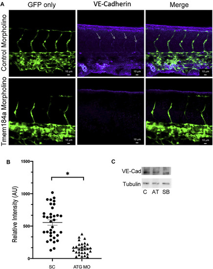
KD of Tmem184a results in decreased levels of VE-cadherin. Embryos were injected with control or ATG-MO for Tmem184a, sorted for GFP positive and fixed at 43 hpf. Embryos were stained with the Anti-zebrafish VE-cadherin antibody as described in Methods and imaged on the Zeiss LSM 880 confocal microscope at 20x. (A) Sections in the mid-section of representative embryos are illustrated. Sections illustrated are maximum intensity images as in Methods. Data from 34 Tmem184a KD ISVs and 29 control ISVs from four embryos of each type were analyzed for VE-cadherin intensity across all ISVs in the mid-sections imaged and a graph illustrating relative VE-cadherin is shown in (B). A student’s T test was used to determine significance, p < 0.001 (C) Western blots of identically treated embryo lysates are shown.
|