
Fig. 6
- ID
- ZDB-FIG-220913-6
- Publication
- Shrestha et al., 2022 - Embryonic Hyperglycemia Delays the Development of Retinal Synapses in a Zebrafish Model
- Other Figures
- All Figure Page
- Back to All Figure Page
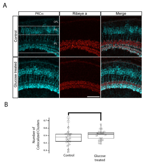
IPL ribbon synapses of bipolar cells are preserved in hyperglycemic larvae. (A) Transverse retinal sections from 5 dpf control (top) and hyperglycemic (bottom) larvae were double immunostained with fluorescently labeled antibodies specific for the rod bipolar cell marker PKCα (cyan; left panels) or ribeye a (red; middle panels); also shown is the overlay of PKCα and ribeye a labeling (merge; right panels). Maximal intensity projections are shown, and the relative positions of the OPL, INL, and IPL are indicated. Scale bar, 5 µm. The expression pattern of PKCα-labeled rod bipolar cell neurons, synaptic ribbons in the IPL layer, and the number of ribbon synapses in rod bipolar cells are normal in 5 dpf hyperglycemic larvae. (B) Quantitative analyses of IHC for colocalization of PKCα and ribeye a are indicated by box-and-whisker plots. Boxes indicate median values, and whiskers indicate 5th–95th percentile values of pooled data from 56 larvae. The data represent the total ribeye a labeling in PKCα-labeled terminals (p > 0.05). Abbreviations used: Dpf, days post-fertilization; PKCα, protein kinase C alpha; SEM, standard error of the mean. |