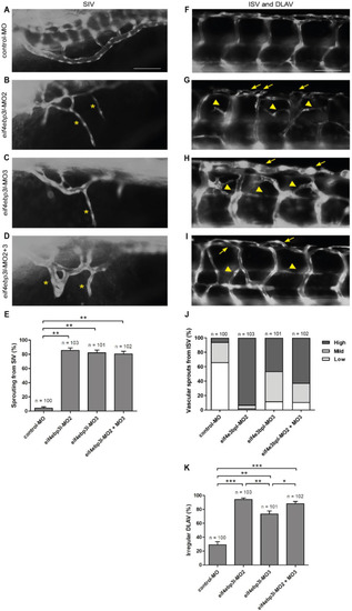
Loss of eukaryotic initiation factor 4E binding protein 3 like (eif4ebp3l) induced sprouting angiogenesis during zebrafish vascular development. (A–I) Lateral view of morpholino-injected Tg(fli1:eGFP) zebrafish at 94 h post fertilization (hpf). Anterior is to the left, abdominal (A–D) or trunk (F–I) regions are shown. Embryos were injected at 1–2 cell stage with control-MO (A), eif4ebp3l-morpholino 2 (MO2), eif4ebp3l-morpholino 3 (MO3) or a combination of the two. (B–D) In eif4ebp3l morphants the sub intestinal vein (SIV) was irregular and formed ventral additional vascular sprouts (yellow starlets). (E) Quantification of the formation of additional vascular sprouts from the SIV. (F–I) Eif4ebp3l morphants displayed vascular sprouting from the intersegmental vessels (ISVs) and within the dorsal longitudinal anastonomic vessel (DLAV). The additional sprouts formed cross links between neighboring ISVs (yellow arrowheads) and led to an increase of width in the DLAV (yellow arrows). (J) Quantification of the number of vascular sprouts of the ISV. Embryos were grouped in low phenotypes (one to two sprouts per ISV), mild (three to four sprouts per ISV) or high phenotypes (>five sprouts per ISV). (K) Quantification of Tg(fli1:eGFP) embryos with irregular DLAV. Comparison between the groups was made by using the Fisher‘s Exact test. p-value adjustments for multiple testing were performed by using the Bonferroni-Holm correction method. Values are mean ± SEM. * refers to p < 0.05, ** to p < 0.005 and *** to p < 0.0005. Scale bar = 50 µm.
|
