FIGURE

Figure 16

ID
ZDB-FIG-220829-114
Publication
Copeland et al., 2022 - Investigating the Transient Regenerative Potential of Cardiac Muscle Using a Neonatal Pig Partial Apical Resection Model
Other Figures
All Figure Page
Back to All Figure Page
Figure 16

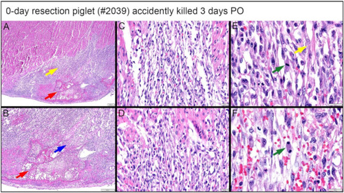
Histological images stained with H & E for the 0-day piglet (#2039) that was accidentally killed by the mother sow three days post apex resection surgery. (A) Image of 0-day piglet heart at 2× magnification; (B) Image of 0-day piglet heart at 4× magnification; the yellow arrow is the border zone of the dense cellular population; the red arrow is fibrin zone/fibrin; the blue arrow is necrotic cardiomyocytes; scale bar = 200 μm. (C) Image of 0-day piglet heart at 20× magnification; (D) Image of 0-day piglet heart at 20× magnification; scale bar = 20 μm. (E) Image of 0-day piglet heart at 40× magnification; (F) Image of 0-day piglet heart at 40× magnification; the green arrow is cells with mitotic events; the yellow arrow indicates the cells resembling immature cardiomyocytes; scale bar = 20 μm. H & E: pink–cytoplasm; blue–nuclei; intense red–erythrocytes.

Expression Data

Expression Detail
Antibody Labeling
Phenotype Data

Phenotype Detail
Acknowledgments
This image is the copyrighted work of the attributed author or publisher, and ZFIN has permission only to display this image to its users. Additional permissions should be obtained from the applicable author or publisher of the image. Full text @ Bioengineering (Basel)