
Figure 12
- ID
- ZDB-FIG-220829-110
- Publication
- Copeland et al., 2022 - Investigating the Transient Regenerative Potential of Cardiac Muscle Using a Neonatal Pig Partial Apical Resection Model
- Other Figures
- All Figure Page
- Back to All Figure Page
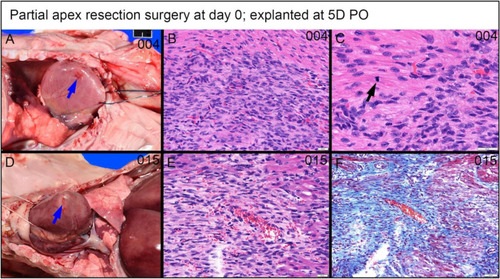
Partial apex resection surgery at 0 days and heart explanted at 5D PO ( |