
- Title
-
Investigating the Transient Regenerative Potential of Cardiac Muscle Using a Neonatal Pig Partial Apical Resection Model
- Authors
- Copeland, K.M., Brazile, B.L., Butler, J.R., Cooley, J., Brinkman-Ferguson, E., Claude, A., Lin, S., Rais-Rohani, S., Welch, B., McMahan, S.R., Nguyen, K.T., Hong, Y., Ramaswamy, S., Liu, Z.P., Bajona, P., Peltz, M., Liao, J.
- Source
- Full text @ Bioengineering (Basel)

( |

Average ejection fraction (percentage) at different time points for each group. ( |
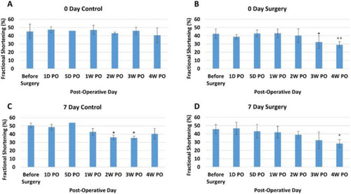
Average fractional shortening (percentage) at different time points for each group. ( |
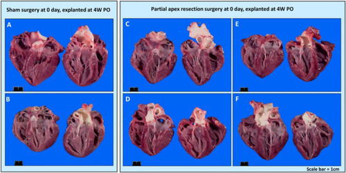
Gross anatomy photos. ( |
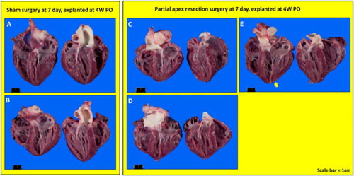
Gross anatomy photos. ( |

Partial apex resection surgery at 0 days and heart explanted at 4W PO-Masson’s Trichrome histology of 0-day control hearts and 0-day surgery hearts. Heart explants were obtained four weeks post-operation (4W PO). ( |
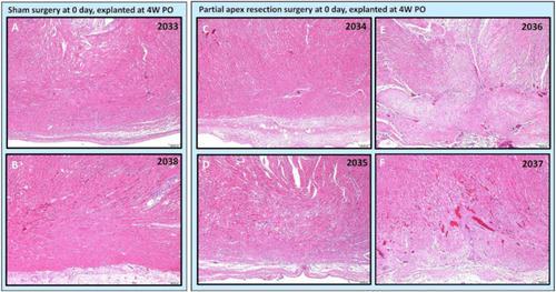
Partial apex resection surgery at 0 days and heart explanted at 4W PO-Hematoxylin and Eosin (H & E) histology of 0-day control hearts and 0-day surgery hearts. Heart explants were obtained four weeks post-operation (4W PO). ( |
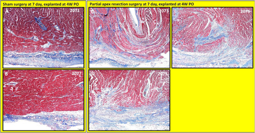
Partial apex resection surgery at seven days and heart explanted at 4W PO-Masson’s Trichrome histology of seven-day control hearts and seven-day surgery hearts. Heart explants were obtained four weeks post-operation (4W PO). ( |
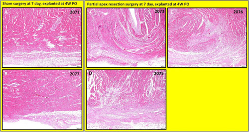
Partial apex resection surgery at seven days and heart explanted at 4W PO-Hematoxylin and Eosin (H & E) histology of seven-day control hearts and seven-day surgery hearts. Heart explants were obtained four weeks post-operation (4W PO). ( |

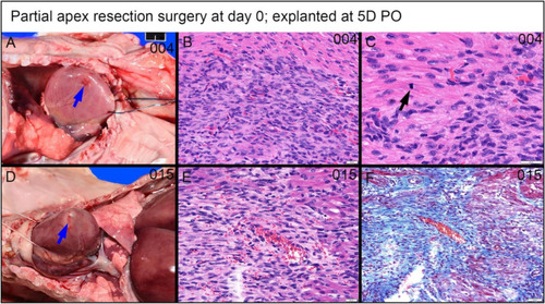
Partial apex resection surgery at 0 day and heart explants at 5D PO and 1W PO-Masson’s Trichrome histology of 0-day surgery heart explants obtained five days post-operation (5D PO): ( |

Partial apex resection surgery at 0 day and heart explants at 5D PO and 1W PO-Hematoxylin and Eosin (H & E) histology of 0-day surgery heart explants obtained five days post-operation (5D PO): ( |
Partial apex resection surgery at 0 days and heart explanted at 5D PO ( |
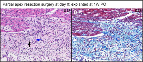
Partial apex resection surgery at 0 days and heart explanted at 1W PO-( |
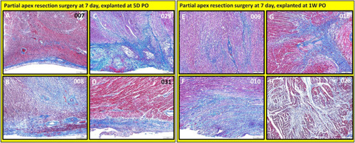
Partial apex resection surgery at seven days and heart explants at 5D PO and 1W PO-Masson’s Trichrome histology of seven-day surgery heart explants obtained five days post-operation (5D PO): ( |
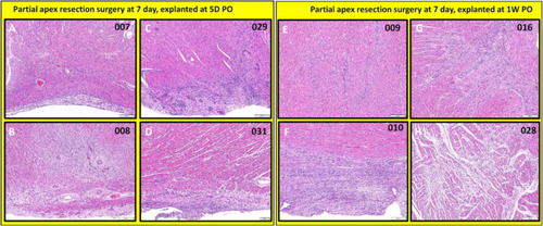
Partial apex resection surgery at seven days and heart explants at 5D PO and 1W PO-Hematoxylin and Eosin (H & E) histology of seven-day surgery heart explants obtained five days post-operation (5D PO): ( |
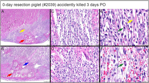
Histological images stained with H & E for the 0-day piglet (#2039) that was accidentally killed by the mother sow three days post apex resection surgery. ( |