
Figure 1.
- ID
- ZDB-FIG-220717-64
- Publication
- Mahony et al., 2021 - Hapln1b, a central organizer of the extracellular matrix, modulates kit signalling to control developmental haematopoiesis
- Other Figures
- All Figure Page
- Back to All Figure Page
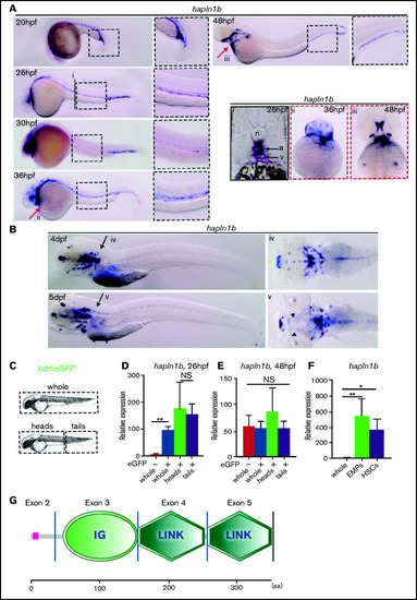
Hapln1b is expressed by vascular cells and hematopoietic progenitors. (A) WISH of hapln1b expression from 20 to 48 hpf. (Ai) Section at 26 hpf, displaying aorta (a) and vein (v) and notochord (n). (Aii-iii) ventral view at 36 and 48 hpf. (B) WISH of hapln1b expression at 4 and 5 dpf. (Biv-v) Dorsal view at 4 and 5 dpf. (C-E) Experimental outline and qPCR expression of hapln1b from FACS-sorted endothelial cells. (F) qPCR expression of hapln1b in FACS-sorted hematopoietic progenitors (EMPs and HSPCs). All qPCR data are from biological triplicates (except for whole GFP− in panel D, where data represent biological duplicates). Data indicate expression relative to ef1a (calculated by [2Ct(hapln1b) − Ct(ef1a)] × 10 000). (D) Two-tailed Student t-test, whole GFP− and GFP+; P = .0035; heads and tails, P = .75. (E-F) Analysis was performed by 1-way analysis of variance with multiple comparisons. (E) Whole GFP− and GFP+, P = .99; whole GFP− and heads, P = .44; and whole GFP− and tails, P = .99. (F) Whole and EMPs, P = .0081; whole and HSPCs, P = .0449. (G) SMART (http://smart.embl.de/) prediction of hapln1b protein structure. IG, immunoglobulinlike domain; Link, HA Link domain; aa, amino acid. |