
- Title
-
Hapln1b, a central organizer of the extracellular matrix, modulates kit signalling to control developmental haematopoiesis
- Authors
- Mahony, C.B., Cacialli, P., Pasche, C., Monteiro, R., Savvides, S.N., Bertrand, J.Y.
- Source
- Full text @ Blood Adv
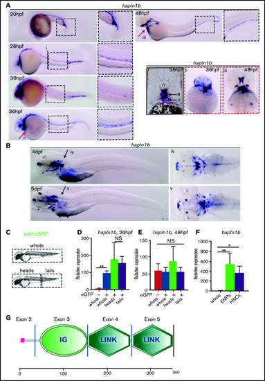
Hapln1b is expressed by vascular cells and hematopoietic progenitors. (A) WISH of hapln1b expression from 20 to 48 hpf. (Ai) Section at 26 hpf, displaying aorta (a) and vein (v) and notochord (n). (Aii-iii) ventral view at 36 and 48 hpf. (B) WISH of hapln1b expression at 4 and 5 dpf. (Biv-v) Dorsal view at 4 and 5 dpf. (C-E) Experimental outline and qPCR expression of hapln1b from FACS-sorted endothelial cells. (F) qPCR expression of hapln1b in FACS-sorted hematopoietic progenitors (EMPs and HSPCs). All qPCR data are from biological triplicates (except for whole GFP− in panel D, where data represent biological duplicates). Data indicate expression relative to ef1a (calculated by [2Ct(hapln1b) − Ct(ef1a)] × 10 000). (D) Two-tailed Student t-test, whole GFP− and GFP+; P = .0035; heads and tails, P = .75. (E-F) Analysis was performed by 1-way analysis of variance with multiple comparisons. (E) Whole GFP− and GFP+, P = .99; whole GFP− and heads, P = .44; and whole GFP− and tails, P = .99. (F) Whole and EMPs, P = .0081; whole and HSPCs, P = .0449. (G) SMART (http://smart.embl.de/) prediction of hapln1b protein structure. IG, immunoglobulinlike domain; Link, HA Link domain; aa, amino acid. |
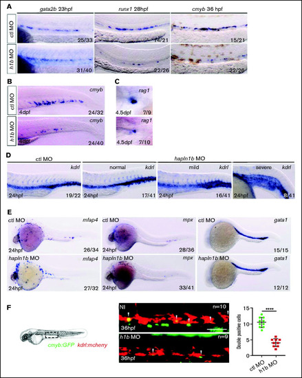
Hapln1b is necessary for the specification of HSPC from the hemogenic endothelium. (A) Gata2b, runx1, and cmyb ISH in control MO, or hapln1b MO-injected embryos (injected at 3 ng throughout, unless otherwise stated). (B) ISH expression of cmyb, in control MO–, or hapln1b MO–injected embryos. (C) Rag1 expression in the thymus after control MO or hapln1b MO injection. (D) ISH expression of kdrl in control MO– or hapln1b MO–injected embryos. Hapln1b morphants displayed 3 phenotypes: normal, mild, and severe. (E) ISH expression of mfap4, mpx, and gata1 in control MO– or hapln1b MO–injected embryos. H1b, hapln1b. (F) Imaging double-transgenic kdrl:mCherry/cmyb:GFP embryos at 36 hpf. Bar represents 50 µm. Control (ctl) MO vs hapln1b mRNA injected, P < .0001. Student unpaired t-test. |

Hapln1b overexpression inhibits HSPC budding and development. (A) ISH expression of kdrl, runx1, and cmyb in non-injected or hapln1b mRNA–injected embryos. (B-C) ISH expression of cmyb in non-injected or hapln1b mRNA–injected embryos. (D-D′) Double-transgenic kdrl:mCherry/cmyb:GFP embryos at 36 and 48 hpf. Bar represents 50 µm. (D′) NI vs +h1b-injected at 36 hpf, P < .0001; and at 48 hpf, P = .0003. (E-E′) Imaging double-transgenic kdrl:mCherry/cmyb:GFP embryos at 3 and 4 dpf. Bar represents 50 µm. (E′) NI vs +h1b injected at 3 dpf, P = .0003; and at 4 dpf, P = .004. NI, non-injected, +h1b: hapln1b mRNA–injected. |

Hapln1b interacts with kitlgb to maintain kitlgb-kitb interactions in the AGM. (A) ISH expression pattern of runx1 in control morphants, kitlga mRNA–injected embryos, kitlgb mRNA–injected embryos, hapln1b morphants, embryos injected with both hapln1b MO and kitlga mRNA, and embryos injected with both hapln1b MO and kitlgb mRNA. (A′) Analysis of runx1 expression. All results are compared with control MO. Data were analyzed by Fisher’s exact test using R. Control (ctl) MO vs +kitlga, P = .05826; NI vs +kitlgb, P = .7939; NI vs +hapln1b, P < .0001; NI vs +hapln1b+kitlga, P < .0001; and NI vs +hapln1b+kitgb: P < .0001. (B) ISH expression of cmyb in NI control embryos, hapln1b mRNA–injected embryos, kitlgb mRNA–injected embryos, and embryos injected with both hapln1b and kitlgb. (B′) analysis of cmyb expression. All analysis is compared with the NI control. Data were analyzed by Fisher’s exact test using R. NI vs +kitlga, P = .8373; NI vs +kitlgb, P = .99; NI vs +hapln1b, P = .0051; NI vs +hapln1b+kitlga, P = .036; and NI vs +hapln1b+kitgb, P = .8617. h1b, hapln1b; ka, kitlga; Kb, kitlgb. (C) Electrostatic potential of KITL, kiltga and kitlgb at biological pH. Blue, positive charge; white, neutral charge; red, negative charge. NI, non-injected. |
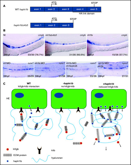
Hapln1b overexpression inhibits cmyb through interactions mediated by the link domains. (A) Schematic representation of WT hapln1b and a truncated version that lacks exons 4 and 5 (hapln1bΔ4Δ5). (B) ISH expression of cmyb in NI control embryos, hapln1b mRNA–injected embryos and hapln1bΔ4Δ5-injected embryos at 36 hpf. ISH expression of runx1 in control MO (ctl MO), hapln1b MO (h1b MO), hapl1b MO, and hapln1bΔ4Δ5 mRNA–injected (h1b MO and h1bΔ4Δ5), and hapl1b MO and hapln1b full-length mRNA (h1bMO and h1b)–injected embryos at 28 hpf. (C) Summary of proposed model. In the wild-type situation, hapn1b helps kitlgb to interact with kitb in the AGM to permit runx1 expression and HSPC formation. Loss of hapln1b results in a loss of kitlgb interaction with kitb, no runx1 expression, and no HSPCs forming. Overexpression of hapln1b results in a dense ECM and reduced kitlgb interaction with kitb, impairing HSPC emergence and survival. HE, hemogenic endothelium; NI, non-injected. |