
Fig. 1
- ID
- ZDB-FIG-220714-2
- Publication
- Kuromiya et al., 2022 - Calcium sparks enhance the tissue fluidity within epithelial layers and promote apical extrusion of transformed cells
- Other Figures
- All Figure Page
- Back to All Figure Page
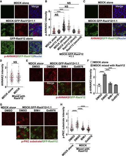
Figure 1. Phosphorylation of AHNAK2 is up-regulated in normal cells mix cultured with RasV12-transformed cells (A and B) Phosphorylation of AHNAK2 in monoculture or mix culture of normal and RasV12-transformed cells. (A) Immunofluorescence images of p-AHNAK2. (B) Quantification of fluorescence intensity of p-AHNAK2; “direct” or “second” indicates cells directly contacting or in the second row. Values are expressed as a ratio relative to the average of MDCK alone. Cumulative data from 3 independent experiments are shown as means ± SDs. ∗∗∗∗p < 0.0001 and NS, not significant (1-way ANOVA with Tukey’s test); n = 150, 150, 355, 127, 150, and 67 cells. (C and D) The level of AHNAK2 proteins in monoculture of normal cells or mix culture of normal and RasV12-transformed cells. (C) Immunofluorescence images of AHNAK2. (D) Quantification of fluorescence intensity of AHNAK2. Values are expressed as a ratio relative to the average of MDCK alone. Cumulative data from 3 independent experiments are shown as means ± SDs. NS, not significant (unpaired 2-tailed Student’s t test); n = 50 cells for each experiment. (E–H) Effect of the pan-PKC inhibitor BIM-I or the Ca2+-dependent conventional PKC inhibitor Go6976 on the phosphorylation of AHNAK2 (E and F) or the PKC substrate (G and H). (E and G) Immunofluorescence images of p-AHNAK2 (E) or p-PKC substrate (G) in the absence or presence of BIM-I or Go6976. (F and H) Quantification of the fluorescence intensity of p-AHNAK2 (F) or p-PKC substrate (H). Values are expressed as a ratio relative to the average of MDCK alone (DMSO). (F) Data are means ± SDs from 3 independent experiments. (H) Cumulative data from 3 independent experiments are shown as means ± SDs. ∗∗∗∗p < 0.0001 (1-way ANOVA with Dunnett’s test); n = 50 cells for each experiment. (A, C, E, and G) Scale bars, 20 μm. See also Figures S1, S2, and Table S1. |