
Fig. 3
- ID
- ZDB-FIG-220713-35
- Publication
- Zhang et al., 2022 - Investigation Driven by Network Pharmacology on Potential Components and Mechanism of DGS, a Natural Vasoprotective Combination, for the Phytotherapy of Coronary Artery Disease
- Other Figures
- All Figure Page
- Back to All Figure Page
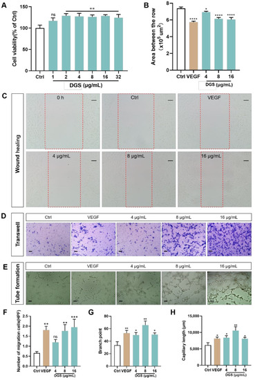
Effect of DGS on HUVECs cells in vitro: (A) An CCK-8 assay was carried out to measure HUVECs viability. (B) Effect of different concentrations of DGS on the migration of HUVECs cells. Results are presented as the mean ± SEM. (C) The healing area of the wound at 0 and 24 h were photographed by microscopy. The red dashed box represents the area counted after migration. Scale bar: 100 μm. (D) The migration of HUVECs in Transwell migration assays. Scale bar: 100 μm. (E) DGS promoted tube formation of HUVECs. Scale bar: 100 μm. (F) Quantification of the number of migrated cells. (G) Quantitative analysis of branch points for tube formation assays. (H) Quantitative analysis of capillary length for tube formation assays. Values are expressed as the mean ± SEM. ns p < 0.05 vs. Control, * p < 0.05 vs. Control, ** p < 0.01 vs. Control, *** p < 0.001 vs. Control, **** p < 0.0001 vs. Control. |