
Fig. 5
- ID
- ZDB-FIG-220622-99
- Publication
- Yang et al., 2022 - Myoneurin regulates BMP signaling by competing with Ppm1a for Smad binding
- Other Figures
- All Figure Page
- Back to All Figure Page
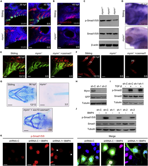
Mynn negatively regulates BMP signaling (A) Expression of dGFP reporter in the pharyngeal region. Mynn−/− mutants and their siblings in Tg(BRE:dGFP) background were stained with the indicated antibodies. Nuclei were counterstained with DAPI (blue). Lateral views, anterior to the left. Scale bars, 20 μm. (B and C) Expression of p-Smad1/5/9 was dramatically reduced in mynn−/− mutants. mynn−/− embryos and their siblings were harvested at 48 hpf, and then stained with anti-p-Smad1/5/9 antibodies (B). Scale bars, 20 μm. The levels of p-Smad1/5/9 in cell lysis of head region were further analyzed by Western blots (C). (D) Expression levels of BMP target gene id1 were detected by in situ hybridization. Embryos were lateral views with anterior to the left. (E) Reactivation of BMP signaling can restore the proliferation of CNCCs in mynn mutant embryos. mynn−/− embryos in Tg(fli1:EGFP) background were injected with or without 10 pg mRNA encoding constitutively active Smad1 protein (caSmad1) at the one-cell stage, and incubated with BrdU at 34 hpf, then harvested at 36 hpf for staining with anti-BrdU (red) and anti-GFP (green) antibodies. Scale bars, 20 μm. (F) Reactivation of BMP signaling can partially restore the differentiation of CNCCs in mynn−/− mutants. mynn−/− embryos were injected with or without 10 pg casmad1 mRNA at the one-cell stage and then immunostained with anti-Col2 antibody at 72 hpf. pq, palatoquadrate; ch, ceratohyal. Scale bars, 20 μm. (G) Alcian blue staining on embryos from different genotypes. Note that the missing cartilages in mynn−/− mutants could be rescued by specific overexpression of casmad1 in sox10+ NCCs. Scale bars, 200 μm. (H) The effectiveness of Mynn shRNAs. HEK293T cells were transfected with the indicated shRNA plasmids and harvested 48 h after transfection for Western blot analyses. (I and J) HEK293 cells transfected with indicated shRNA plasmids were treated with TGF-β1 (5 ng/mL) for 2 h (I) or BMP4 (20 ng/mL) for 4 h (J), and then collected for Western blots with the indicated antibodies. (K) Hep3B cells transfected with plasmids expressing indicated shRNAs and GFP proteins were treated with or without BMP4 for 4 h. The expression levels of p-Smad1/5/9 were determined with immunostaining. As indicated with white arrowheads, the expression of p-Smad1/5/9 was obviously decreased in the cells expressing Mynn shRNAs. Scale bars, 20 μm. |
| Gene: | |
|---|---|
| Antibodies: | |
| Fish: | |
| Anatomical Terms: | |
| Stage Range: | Prim-25 to Pec-fin |