
Fig. 1
- ID
- ZDB-FIG-220622-95
- Publication
- Yang et al., 2022 - Myoneurin regulates BMP signaling by competing with Ppm1a for Smad binding
- Other Figures
- All Figure Page
- Back to All Figure Page
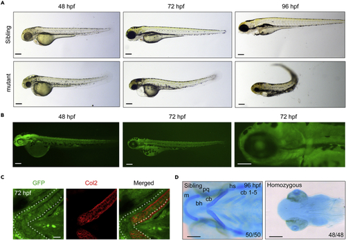
The GFP expression and developmental defects in T054 embryos (A) Morphology of T054 mutants at indicated stages. Note that mutant embryos had a darker head, rough skin, reduced size of head and eyes, severe craniofacial jaw malformations, and curly posterior trunk with stage-dependent degrees. Scale bars, 200 μm. (B) Fluorescent images of T054 embryos. The pictured embryos were heterozygotes. Scale bars, 200 μm. (C) Immunostaining of chondrocytes by anti-Col2 and GFP antibody. T054 heterozygous embryos at 72 hpf were co-immunostained with anti-Col2 (red) and anti-GFP (green) antibodies. Scale bar, 20 μm. (D) Craniofacial cartilages stained with Alcian blue. Note that almost no cartilages could be seen in the T054 mutant embryos. The cartilages were positioned with anterior to the left, and ceratobranchial (cb), ceratohyal (ch), Meckel’s cartilage (m), palatoquadrate (pq), and hyosymplectic (hs) cartilages were shown. Scale bars, 200 μm. |
| Antibodies: | |
|---|---|
| Fish: | |
| Anatomical Term: | |
| Stage Range: | Long-pec to Protruding-mouth |
| Fish: | |
|---|---|
| Observed In: | |
| Stage Range: | Long-pec to Day 4 |