
FIGURE
Fig. 3
- ID
- ZDB-FIG-220621-13
- Publication
- Liu et al., 2022 - LAPTM4B-35 promotes cancer cell migration via stimulating integrin beta1 recycling and focal adhesion dynamics
- Other Figures
- All Figure Page
- Back to All Figure Page
Fig. 3
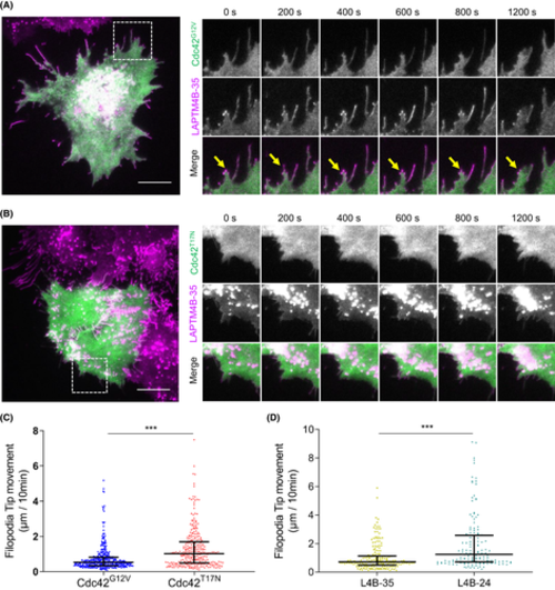
LAPTM4B-35 distributing on Cdc42-induced filopodia. A, A431 cells stably expressing LAPTM4B-35-mCherry were transiently transfected with constitutively active Cdc42G12V-GFP and imaged by live total internal reflection fluorescence (TIRF) microscopy. Scale bar: 10 μm. A magnified region of interest at different time points is visible on the right. Yellow arrows highlight LAPTM4B-35 positive filopodia. B, A431 cells stably expressing LAPTM4B-35-mCherry were transiently transfected with dominant negative Cdc42T17N-GFP, and imaged by TIRF microscopy. Scale bar: 10 μm. A region of interest at different time points is on the right. C, A431 cells stably expressing LAPTM4B-35-mCherry were transiently transfected with either constitutively active Cdc42G12V-GFP or dominant negative Cdc42T17N-GFP and imaged by TIRF microscopy. The distance of filopodia tip movement in 10 min was quantified. Each dot represents a single measurement, middle line represents mean, and whiskers represent interquartile range. D, Cells stably expressing Flag-tagged LAPTM4B-35 and LAPTM4B-24 were transfected with integrin beta1-mCherry to visualize cell protrusions, and cells were imaged by live TIRF microscopy. The distance of filopodia tip movement in 10 min was quantified. Data are from three independent experiments and >10 videos per condition. LAPTM4B-35: n = 196 filopodia; LAPTM4B-24: n = 151 filopodia; p = 8.8 × 10⁻⁸. Each dot represents a single measurement, middle line represents mean, and whiskers represent interquartile range
|
Expression Data
Expression Detail
Antibody Labeling
Phenotype Data
Phenotype Detail
Acknowledgments
This image is the copyrighted work of the attributed author or publisher, and
ZFIN has permission only to display this image to its users.
Additional permissions should be obtained from the applicable author or publisher of the image.
Full text @ Cancer Sci.