|
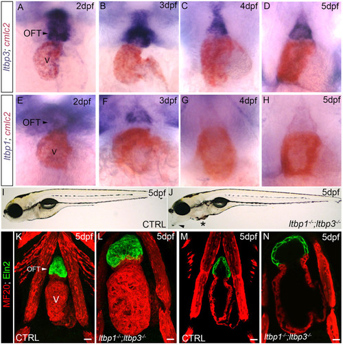
ltbp1, ltbp3 DKO zebrafish exhibit OFT aneurysm and ventricular dilation. (A-H) Brightfield images of hearts in wild-type zebrafish at 2 dpf (A,E), 3 dpf (B,F), 4 dpf (C,G), and 5 dpf (D,H) processed for double whole-mount in situ hybridization to detect ltbp3 (A-D, blue signal) or ltbp1 (E-H, blue signal) transcripts and the myocardial transcript cmlc2 (myl7; A-H; red signal). Little to no variation was observed between animals within each group (n>10/group). (I,J) Brightfield images of 5 dpf control (CTRL; I) and ltbp1−/−; ltbp3−/− (J) larvae. Arrowhead and * in J highlight jaw protrusion and mild pericardial edema, respectively, observed in double mutants. (K-N) Confocal images of hearts in 5 dpf CTRL (K,M) and ltbp1−/−; ltbp3−/− (L,N) larvae double immunostained for striated muscle (MF20, red) and Eln2-positive OFT smooth muscle (green). The single optical sections shown in M and N were taken from the images shown in K and L. V, ventricle. Scale bars: 20 µm.
|