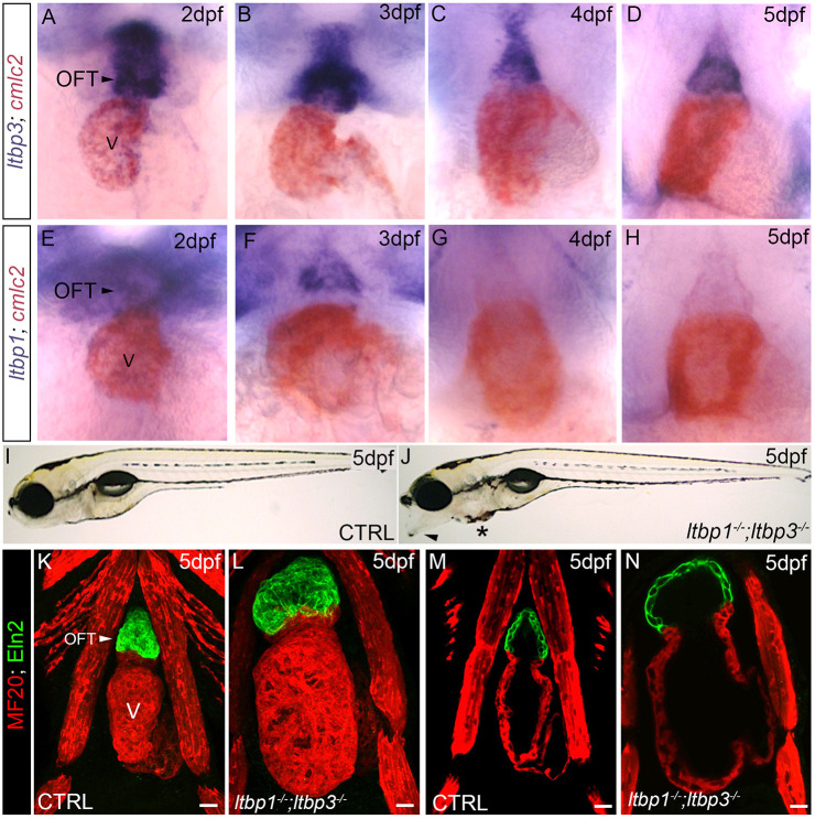
ltbp1, ltbp3 DKO zebrafish exhibit OFT aneurysm and ventricular dilation. (A-H) Brightfield images of hearts in wild-type zebrafish at 2 dpf (A,E), 3 dpf (B,F), 4 dpf (C,G), and 5 dpf (D,H) processed for double whole-mount in situ hybridization to detect ltbp3 (A-D, blue signal) or ltbp1 (E-H, blue signal) transcripts and the myocardial transcript cmlc2 (myl7; A-H; red signal). Little to no variation was observed between animals within each group (n>10/group). (I,J) Brightfield images of 5 dpf control (CTRL; I) and ltbp1−/−; ltbp3−/− (J) larvae. Arrowhead and * in J highlight jaw protrusion and mild pericardial edema, respectively, observed in double mutants. (K-N) Confocal images of hearts in 5 dpf CTRL (K,M) and ltbp1−/−; ltbp3−/− (L,N) larvae double immunostained for striated muscle (MF20, red) and Eln2-positive OFT smooth muscle (green). The single optical sections shown in M and N were taken from the images shown in K and L. V, ventricle. Scale bars: 20 µm.
This image is the copyrighted work of the attributed author or publisher, and
ZFIN has permission only to display this image to its users.
Additional permissions should be obtained from the applicable author or publisher of the image.
Full text @ Dis. Model. Mech.
Your Input Welcome
Thank you for submitting comments. Your input has been emailed to ZFIN curators who may contact you if
additional information is required.
Oops. Something went wrong. Please try again later.