
FIGURE 7
- ID
- ZDB-FIG-220302-92
- Publication
- Babakhanova et al., 2021 - Rapid Directed Molecular Evolution of Fluorescent Proteins in Mammalian Cells
- Other Figures
- All Figure Page
- Back to All Figure Page
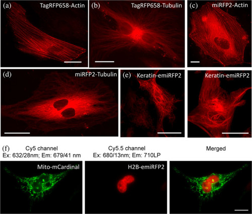
Fluorescence imaging of TagRFP658, miRFP2, and emiRFP2 fusions expressed in HeLa cells. (a–e) Representative fluorescence images of live HeLa cells transfected with (a) TagRFP658-α-Tubulin (n = 11 cells from two independent transfections) and (b) TagRFP658-β-actin (n = 19 cells from two independent transfections). (c) miRFP2-α-Tubulin (n = 15 cells from two independent transfections), (d) miRFP2-β-actin (n = 21 cells from two independent transfections), and (e) Keratin-emiRFP2 (n = 8 cells from two independent transfections). Imaging conditions: (a–d) excitation 631/28 nm from an LED, emission 664LP under wide-field microscope. (e) Excitation 640 nm from laser, emission 670–750 nm under confocal microscope. (f) Dual-color near-infrared (NIR) imaging of live HeLa cells co-expressing Mito-mCardinal and H2B-emiRFP2 (n = 12 cells from two independent transfection) |