
Fig. 3
- ID
- ZDB-FIG-220301-9
- Publication
- Donati et al., 2021 - Planar polarization of cilia in the zebrafish floor-plate involves Par3-mediated posterior localization of highly motile basal bodies
- Other Figures
- All Figure Page
- Back to All Figure Page
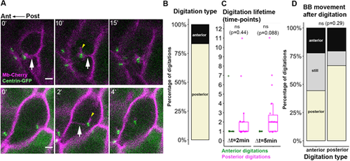
Membrane digitations link BBs to ant/post membranes during FP polarization. (A) Images from live imaging (Δt=2 or 5 min) showing posterior (top) and anterior (bottom) digitations (white arrows). Time (in min) is indicated in the upper-left corner. Short mCherry-positive digitations, presumably cilia, were associated with the BB in some cases (yellow arrowheads). (B) Percentage of posterior and anterior digitations from live-imaging analyses (54 digitations, eight embryos, 22 cells). (C) Number of time-points at which anterior or posterior digitations were detected in movies with Δt=2 or 5 min (Δt2 min: 6 anterior, 23 posterior digitations, four embryos, eight cells. Δt5 min: three anterior and 22 posterior digitations, six embryos, 13 cells). ns, P>0.05 (Wilcoxon rank sum test). Digitations were most often observed in a single or two consecutive timeframes. Box boundaries represent the first and third quartiles of the distribution, and boxplot whiskers span 1.5 times the interquartile range of the distribution. (D) BB movements after anterior or posterior digitation (48 digitations, eight embryos, 22 cells). ns, P>0.05 (Fisher's exact test). Scale bars: 2 µm. |