
Fig. 3
- ID
- ZDB-FIG-220224-37
- Publication
- Paramos-de-Carvalho et al., 2021 - Targeting senescent cells improves functional recovery after spinal cord injury
- Other Figures
- All Figure Page
- Back to All Figure Page
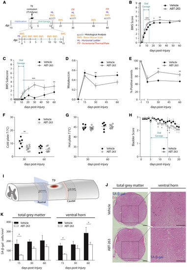
Targeting senescent cells with ABT-263 improves motor, sensory, and bladder function recovery following a spinal cord injury in mice (A) Schematic of the experimental setup. Animals were habituated to the different behavioral setups for a 15-day period, before being submitted to a moderate-to-severe (force, 75 Kdyne; displacement, 550–750 μm) T9 contusion injury. Injured animals received daily vehicle or ABT-263 via oral gavage, from 5 to 14 dpi. (B and C) Basso Mouse Scale (BMS) score and subscore were evaluated in an open field at different time-points (0, 1, 3, 5, 7, 10, 12, 15, 21, 30, 45, and 60 dpi). n = 18–19. (D and E) The locomotor performance in the horizontal ladder (HL) was assessed at −1 (control), 15, 30, and 60 dpi by quantifying the total number of mistakes per centimeter and the percentage of singular positive events (plantar step, toe step, and skip) measured and averaged across three successful trials. n = 3-6. (F and G) Thermal allodynia was tested at 30 and 60 dpi by determining the temperature at which injured mice reacted to a cold or hot stimulus. n = 6–8. (H) Bladder function was grossly evaluated by attributing a bladder score to the amount of urine collected each time a bladder was manually voided. n = 18–19. (I) SA-β-gal+ cells were quantified in a total of 10 different transversal sections (5 rostral and 5 caudal) along 2.0 mm at the lesion periphery. A 0.5-mm interval (red dashed zone) was established between the lesion and the beginning of the quantification region. (J) Eosin counterstaining was performed after cryosectioning. SA-β-gal+ cells (blue) were quantified in the total sectional gray matter and only at the ventral horn. Scale bars, 200 μm. (K) Quantifications were performed at all experimental endpoints (15, 30, and 60 dpi). At 15 dpi, ABT-263 treatment significantly decreased the number of SA-β-gal+ cells/mm2 in the total gray matter and in the ventral horn by 68.4% and 58.0%, respectively. At 60 dpi, a significant reduction (41.9%) of SA-β-gal+ cells/mm2 in ABT-263-treated animals was still observed in the ventral horn. n (15 dpi) = 3–4; n (30 dpi) = 3–4; n (60 dpi) = 2–3. Data are presented as mean ± SEM. ∗p < 0.05, ∗∗p < 0.01, ∗∗∗p < 0.001, ABT-263 versus vehicle. |