
Fig. 6
- ID
- ZDB-FIG-220107-25
- Publication
- Meseguer-Ripolles et al., 2021 - Dimethyl fumarate reduces hepatocyte senescence following paracetamol exposure
- Other Figures
- All Figure Page
- Back to All Figure Page
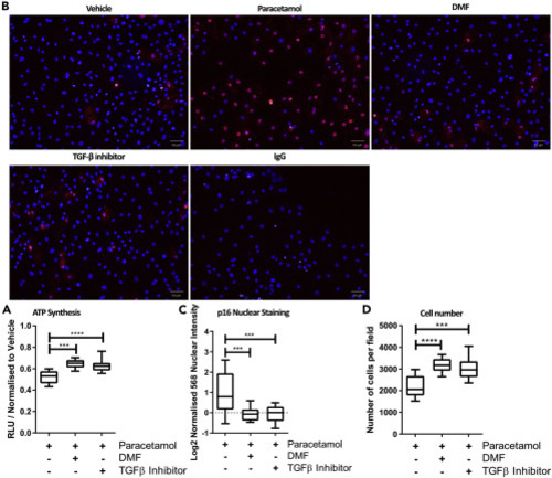
(A) 24 hr treatment of DMF (10 μM) or SB-431542 (10 μM), following 24 hparacetamol (30 mM) exposure, improved ATP levels in HLCs. n = 12. One-way ANOVA test and post-hoc Tukey multiple-comparison test was used. ∗∗p < 0.01, ∗∗∗p < 0.001. Box plot were whiskers represent Min to Max. (B) 6 hr treatment of DMF (10 μM) or SB-431542 (10μM) following 24 hr paracetamol (30 mM) exposure reduces p16 expression and nuclear staining in HLCs. Scale bar represents 50 μm. (C) Single-cell image analysis quantification of p16 shows a significant increase of p16 nuclear intensity when HLCs are treated with paracetamol (30 mM) alone. n = >6. One-way ANOVA test and post-hoc Tukey multiple-comparison test was used. ∗∗∗p < 0.001. Box plot were whiskers represent Min to Max. (D) Treatment with DMF (10 μM) or SB-431542 (10 μM) for 6 hr following 24hr paracetamol (30 mM) exposure for 24 hr significantly prevented cell loss. n = >6. One-way ANOVA test and post-hoc Tukey multiple-comparison test was used. ∗∗∗p < 0.001 ∗∗∗∗p < 0.0001. Scale bar represents 50μm. Box plot were whiskers represent Min to Max. |