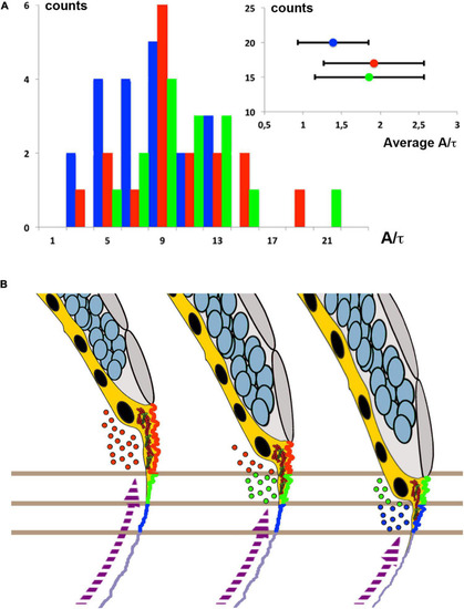
Membrane cortical tension and endocytosis at the E-YSL are necessary for epiboly progression. (A) The distribution (counts) of instant retraction velocities (A/t) after laser surgery of the actomyosin cortex of Myosin-GFP (Tg (β-actin:MYL12.1-eGFP) rab5ab YMOs at 55% epiboly (blue) shows a significant reduction (Wilcoxon test p < 0.01) versus wild type (red) and control YMOs (green). The instant velocity estimate was extracted from the exponential fit of the distance between fronts (see section “MATERIALS AND METHODS”). The averaged instant retraction velocity (A/t) for rab5ab YMOs was 1.39 ± 0.46 μm (n = 20), while control YMOs reach 1.86 ± 0.71 μm (n = 17) and wild type embryos 1.92 ± 0.65 μm (n = 15) (inset). Counts represent the number of analyzed laser cuts for each condition. (B) Proposed model of epiboly progression. The contractile E-YSL and the imbalance of stiffness between the EVL and the yolk cell surface account for epiboly progression. Rab5ab-mediated yolk cell localized endocytosis (color coded dots) accounts for the reduction of the yolk cell surface coupled to the progression of EVL (gray) and DCs (blue) toward the vegetal pole. Membrane removal associates to the convolution and contraction of the E-YSL surface and the recruitment of actin and myosin (purple dashed arrow) from vegetally located pools. Three chronological time points are shown. Different sequential zones on the surface of the E-YSL are color-coded. Actin and myosin are diagrammatically illustrated in red and green within the YSL (yellow).
|
