
Fig 6
- ID
- ZDB-FIG-211116-15
- Publication
- Bühler et al., 2021 - Histone deacetylase 1 controls cardiomyocyte proliferation during embryonic heart development and cardiac regeneration in zebrafish
- Other Figures
- All Figure Page
- Back to All Figure Page
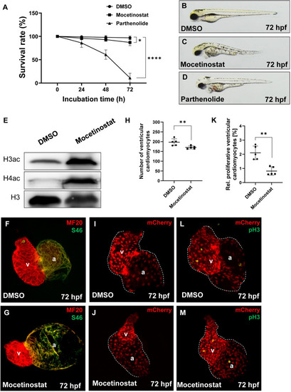
Mocetinostat treatment of zebrafish embryos induces the bal phenotype.
(A) Survival rate of Mocetinostat- and Parthenolide-treated embryos, compared to the DMSO control. At 24 hpf (DMSO: 99.0 ± 2.0%; Mocetinostat: 96.0 ± 3.27%; Parthenolide: 86.0 ± 9.52%, n = 4), at 48 hpf (DMSO: 98.0 ± 4.0%; Mocetinostat: 93.0 ± 3.83%; Parthenolide: 61.0 ± 10.0%, n = 4) and at 72 hpf (DMSO: 97.0 ± 3.83%; Mocetinostat: 87.0 ± 6.83%; Parthenolide: 11.0 ± 10.0%, n = 4). (B-D) Lateral view of inhibitor- or DMSO-treated embryos at 72 hpf. (E) Western blot analysis of acetylation levels of Histone 3 (H3ac) and 4 (H4ac) of lystes from Mocetinostat and DMSO-treated embryos, show a pronounced hyperacetylation of H3 and H4 in Mocetinostat-treated embryos. pan Histone 3 served as a loading control. (F-G) IF staining against meromyosin (MF20) (red) and atrial-specific myosion (S46) (green) demonstrated normal chamber specification of Mocetinostat-treated hearts. (H-J) Number of ventricular cardiomyocytes was reduced in Mocetinostat treated Tg(myl7:mCherry) embryos (DMSO: 197.0 ± 17.39; Mocetinostat: 170.4 ± 7.7, n = 5). (K-M) The rate of ventricular pH3+ cardiomyocytes was reduced in Mocetinostat-treated embryos as well (DMSO: 2.1 ± 0.48%; Mocetinostat: 0.8± 0.30%, n = 5). Error bars indicate s.d., *p < 0.05, **p < 0.01, ****p < 0.0001. |
| Gene: | |
|---|---|
| Antibodies: | |
| Fish: | |
| Condition: | |
| Anatomical Terms: | |
| Stage: | Protruding-mouth |
| Fish: | |
|---|---|
| Conditions: | |
| Observed In: | |
| Stage Range: | Prim-5 to Protruding-mouth |