
Fig 7
- ID
- ZDB-FIG-211116-16
- Publication
- Bühler et al., 2021 - Histone deacetylase 1 controls cardiomyocyte proliferation during embryonic heart development and cardiac regeneration in zebrafish
- Other Figures
- All Figure Page
- Back to All Figure Page
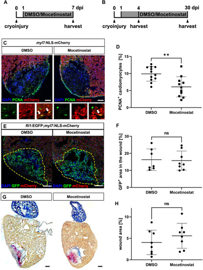
Hdac1 is involved in heart regeneration after cryoinjury.
(A, B) Experimental timeline of cryoinjury, treatment and harvesting of the hearts for the analysis of cardiomyocyte proliferation (A) revascularization (4 dpi) and scar formation (30 dpi) (B). (C, D) Immunohistochemistry for mCherry (red, cardiomyocytes) and PCNA (green) on 7 dpi hearts of Tg(myl7:NLS-mCherry) fish treated with either DMSO or Mocetinostat from 1 to 7 dpi, showed reduced rate of cycling cardiomyocytes at the wound border zone in Mocetinostat-treated hearts (% of PCNA+/mCherry+ cardiomyocytes DMSO 9.91 ± 2.14, n = 12, Mocetinostat 6.1 ± 2.96, n = 11, p = 0.0018). (E, F) Native GFP (green, endothelial cells) and mCherry (red, cardiomyocytes) fluorescence in 4 dpi hearts of Tg(fli:GFPy1;myl7:NLS-mCherry) fish treated with DMSO or Mocetinostat from 12 hpi to 4 dpi, showed no changes in revascularization (% of GFP+ wound area: DMSO Ctrl 16.26 ± 6.34, n = 7, Mocetinostat 15.74 ± 5.62, n = 8, p > 0.9999). (G, H) Acid Fuchsin Orange G (AFOG) staining of 30 dpi hearts treated with DMSO or Mocetinostat from 1 to 30 dpi, have no significant difference in wound size (DMSO 4.05 ± 4.85, n = 8, Mocetinostat 5.59 ± 2.91, n = 8, p = 0.3282). Error bars indicate s.d.; **p < 0.01, ns, not significant. Scale bars, 100 μm. |
| Genes: | |
|---|---|
| Antibody: | |
| Fish: | |
| Conditions: | |
| Anatomical Terms: | |
| Stage: | Adult |
| Fish: | |
|---|---|
| Condition: | |
| Observed In: | |
| Stage: | Adult |