|
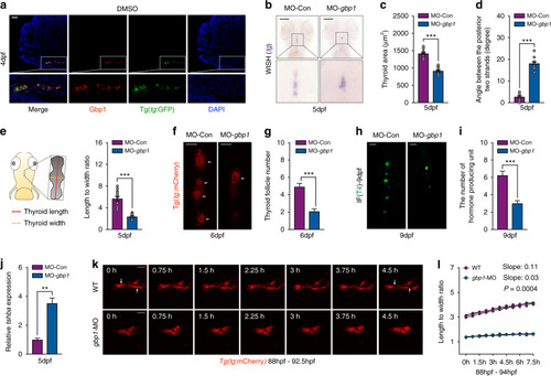
<italic>Gbp1</italic> knockdown led to hypothyroidism and thyroid developmental abnormalities in zebrafish embryos.(a) Representative image showing the expression of Gbp1 in 4-dpf thyroid tissue under Tg(tg:GFP) background using immunofluorescence (IF) assay. (b) Whole-mount in situ hybridization (WISH) assessment of tg expression in gbp1 morphants at 5 dpf. (c–e) Statistical calculation of thyroid area (c), angle between the posterior strands (d), and length to width ratio (e) in gbp1 morphants. N = 12. Left schematic illustration in (e) showing how the length and width of the zebrafish thyroid primordium (TP) is calculated. While the length is measured from the front point to the ending point of the TP along the pharyngeal midline, the width is the measured by the widest axis vertical to the length axis. (f, g) Representative image (f) and statistical analysis (g) of follicle formed with gbp1 knockdown under Tg(tg:mCherry) background. N = 12. Bars = 20 μm. (h,i) Representative image (h) and statistical analysis (i) of T4 producing units with gbp1 knockdown. N = 12. Bars = 20 μm. (j) Tshba transcripts detected in 5-dpf wild-type (WT) and gbp1 morphants. Means ± SEM are shown for three independent experiments. (k) Representative images chosen from Movie S1 and Movie S2 showing the differential growth mode of TP in 88 hpf−92.5 hpf WT and gbp1 deficient embryos. Bars = 20 μm. (l) Statistical calculation of the dynamic changes of the length to width ratio in WT and gbp1 morphants from 88 to 94 hpf. N = 3. The arrows indicate the separation of follicles after 4.5 h morphogenesis. **P < 0.01; ***P < 0.001 (Student’s t-test).
|

