FIGURE

Figure 1

ID
ZDB-FIG-211002-1
Publication
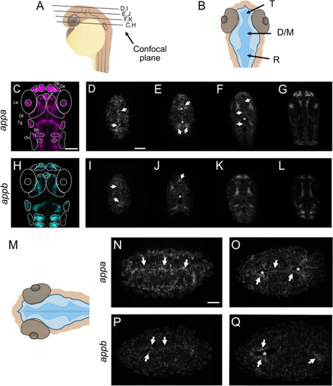
Chebli et al., 2021 - The localization of amyloid precursor protein to ependymal cilia in vertebrates and its role in ciliogenesis and brain development in zebrafish
Other Figures
All Figure Page
Back to All Figure Page
Figure 1

Expression pattern of appa and appb mRNA. (A,B) Schematic representations of head and ventricle morphology of a 30 hpf zebrafish larvae, lateral (A) and dorsal (B) view. (C,H) Whole-mount fluorescent in situ of appa (C) and appb (H) in 30 hpf WT zebrafish larvae. Single focal planes, dorsal to ventral, of whole-mount larvae of appa (DG) and appb (IL) probe. (M) Schematic view of focal plane of the dorsal area of the brain ventricle. (NQ) Single focal plane at high magnification (40×) of appa (N,O) and appb (P,Q) probes. T telencephalic ventricle, D/M diencephalic/mesencephalic ventricle, R rhombencephalic ventricle, Ob olfactory bulb, Oe olfactory epithelium, P pituitary gland, Le lens, Ot optic tectum, Tg trigeminal ganglia, Rh rhombomeres, Ov otic vesicle. Magnification: (CL) = 20×, (NQ) = 40×. Scale bar: (C) = 100 µm, (D) = 50 µm, (N) = 25 µm. *Indicates ventricular space and white arrows highlight appa and appb expression at the ventricular borders.

Expression Data
Genes:
Fish:
Anatomical Terms:
Stage: Prim-15

Expression Detail
Antibody Labeling
Phenotype Data

Phenotype Detail
Acknowledgments
This image is the copyrighted work of the attributed author or publisher, and ZFIN has permission only to display this image to its users. Additional permissions should be obtained from the applicable author or publisher of the image. Full text @ Sci. Rep.