|
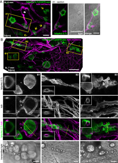
Interaction of early adipocytes with blood vessels. Live larvae from the cross of fabp4a(-2.7):EGFPcaax and kdlr:mCherry fish lines were imaged through confocal microscopy. Images presented here are 3D projections or single sections, as indicated. (A) Larva of SL 6 mm (13 dpf) with many EGFP+ cells in its abdominal area, a few of them having lipid droplets (inset A1). Some of the cells are in contact with blood vessels (double arrows) and some of them are not (single arrows). (B) Larva of SL 7 mm (16 dpf), with PVAT and AVAT depots (only some cells of each depot expresses EGFP). Insets B1, B2 and B3 show EGFP+ cells with lipid droplets in close apposition to blood vessels and in some cases surrounding them (B2). 3D projections and sections through the position indicated by the dashed line are shown. Scale bars: A: 100 μm; B: 100 μm (panoramic view), 20 μm (insets).
|