FIGURE

Figure 5

ID
ZDB-FIG-210822-12
Publication
Holmgren et al., 2021 - Influence of Mpv17 on Hair-Cell Mitochondrial Homeostasis, Synapse Integrity, and Vulnerability to Damage in the Zebrafish Lateral Line
Other Figures
All Figure Page
Back to All Figure Page
Figure 5

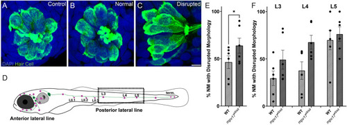
Mechanical overstimulation results in morphological disruption of neuromasts more frequently in mpv17a9/a9 larvae than in WT. (A–C) Maximum-intensity projections of confocal images showing hair cells labeled with Parvalbumin and all nuclei labeled with DAPI in control neuromasts (A) and strong water current exposed neuromasts with normal (B) or disrupted (C) morphology. (D) Schematic of larval zebrafish indicating the placement of neuromasts (pink dots) and afferent nerves (green lines). Neuromasts L3, L4, and L5 (dashed rectangle) were examined in this study. (E,F) Quantification of disrupted neuromasts, both overall (E) and separated by position on the body (F). Each point indicates the percentage of neuromasts with disrupted morphology in a single experimental trial. The frequency of disrupted neuromasts was higher in the more posterior L5 neuromasts; mpv17a9/a9 neuromasts showed disrupted morphology more frequently than WT (*P = 0.0232). n = 9–64 neuromasts (L3, L4, and L5) per trial; N = 6 experimental trials. Scale bar: 5 μm. Error bars: SEM.

Expression Data

Expression Detail
Antibody Labeling
Phenotype Data

Phenotype Detail
Acknowledgments
This image is the copyrighted work of the attributed author or publisher, and ZFIN has permission only to display this image to its users. Additional permissions should be obtained from the applicable author or publisher of the image. Full text @ Front. Cell. Neurosci.