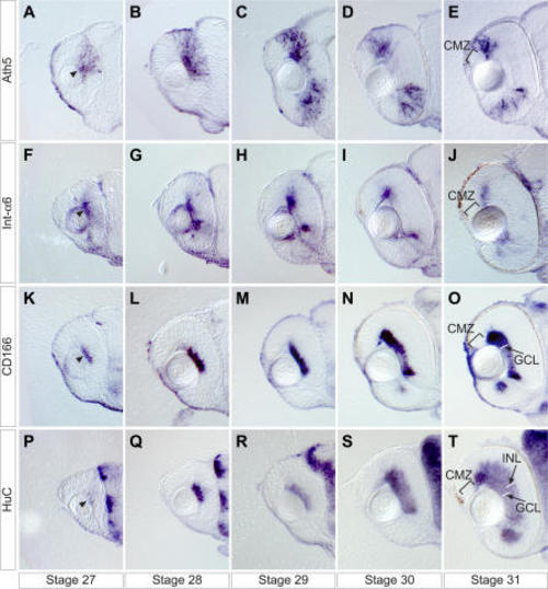
FIGURE

Figure 2

ID
ZDB-FIG-210821-11
Publication
Del Bene et al., 2007 - In vivo validation of a computationally predicted conserved Ath5 target gene set
Other Figures
All Figure Page
Back to All Figure Page
Figure 2

Developmental Time-Course of the Expression of <italic>ath5</italic> (A–E) and Three Examples Representing the Major Groups of Target Genes

Embryos are stained by whole mount in situ hybridization and sectioned transversally at the level of the optic nerve. int-α6 overlaps with ath5 expression and follows it (F–J). CD166 is continuous with the ath5 expression domain and then remains in the cells that had expressed ath5 in the ganglion cell layer (K–O). HuC is expressed in the ganglion cell layer and part of the inner nuclear layer (P–T), during medaka retina development from stage 27 to stage 31. Arrowheads indicate the simultaneous onset of the expression in the central retina of the target genes within ath5 expression domain at stage 27. CMZ, ciliary marginal zone; GCL, ganglion cell layer; and INL, inner nuclear layer.

Expression Data

Expression Detail
Antibody Labeling
Phenotype Data

Phenotype Detail
Acknowledgments
This image is the copyrighted work of the attributed author or publisher, and ZFIN has permission only to display this image to its users. Additional permissions should be obtained from the applicable author or publisher of the image. Full text @ PLoS Genet.