
Figure 1
- ID
- ZDB-FIG-210801-96
- Publication
- Quadri et al., 2021 - Phosphorylation of H3-Thr3 by Haspin Is Required for Primary Cilia Regulation
- Other Figures
- All Figure Page
- Back to All Figure Page
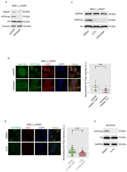
Haspin is active in G0 phase. (a) RPE-1_hTERT cells stably silenced with shSCR (control) or shHaspin were seeded and harvested after 48 h serum starvation to induce entry in G0. Silencing efficiency and H3-T3 phosphorylation were monitored by Western blotting; (b) immunofluorescence against Ki67 (proliferation marker) and H3-T3 in shSCR and shHaspin cells serum-starved for 48 h. Histone phosphorylation was quantified and is reported to the right. Silencing control is shown in Supplementary Figure S1e; (c) RPE-1_hTERT cells were seeded and serum-starved for 48 h, then treated for a further 24 h with DMSO or Haspin inhibitor 5-ITu (10 nM) and CHR-6494 (50 nM); H3T3p was monitored by Western blotting; (d) cells treated with 5-ITu were processed for immunofluorescence against Ki67 and H3-T3. Histone phosphorylation was quantified and is reported to the right; (e) Western blot analysis monitoring the levels of H3-T3p in SH-SY5Y that were differentiated into neuron-like cells, as described in Material and Methods, and then incubated for 24 h with 10 nM 5-ITu. Graphs in b, d show the median abundance of phosphorylated H3-Thr3; boxes include 50% of the data points, notch represent confidence interval (median ±1.58 IQR/sqrt(n)). t-test was applied as a statistical measurement, n.s.; not significant, *** p < 0.005. Scale bars in b, d: 20 µm. |