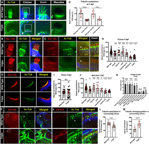
Decrease of acetylation in cilia in hdac mutants. (A) Comparison of levels of tubulin acetylation (green) between 3 dpf wild-type embryos and triple mutant embryos in cristae and macule. Squares shows zoomed in areas. (B–D) Double staining for acetylated (green) and glutamylated (red) tubulin, 5 dpf of wild type and triple mutants. (B) cristae (C) macule, and (D) nose. (E) Increased tubulin acetylation (green) of cell bodies in the retina of 5 dpf embryos, and increased levels of mono-glycylation in a subset of cilia (red). (F) Zoom of connecting cilia increased mono-glycylation as well as a lower level of acetylation in the triple mutants. (G) Level of tubulin acetylation in cilia in wild type and triple mutants, normalized to wild type, for cristae and macule of 3 dpf embryos. (I) Unaffected level of acetylation in nose cilia. Change of cristae (H) macule (J) and cilia tubulin acetylation in hdac6, hdac10, sirt2, hdac6/hdac10, hdac6/sirt2, and triple mutants. (K) Comparison of cilia length between wild type and triple mutants in cristae (anterior and lateral posterior) macule and nose. (L) Decrease in connecting cilium axoneme acetylation in photoreceptors in triple mutants and M) increase in tubulin mono-glycylation in triple mutant cilia. Asterisks show increased level of tubulin acetylation in cell bodies. [Mean with 95% CI. (G,I,L,M)t-test *p < 0.05, **p < 0.01, ***p < 0.005, ****p < 0.0001]. ns, no significant. [(H,J,K) one-way ANOVA *p < 0.05, **p < 0.01, ***p < 0.005, ****p < 0.0001]. ns, no significant.
|
