FIGURE

Figure 6

ID
ZDB-FIG-210628-51
Publication
Ishii et al., 2021 - Correlative microscopy and block-face imaging (CoMBI) method for both paraffin-embedded and frozen specimens
Other Figures
All Figure Page
Back to All Figure Page
Figure 6

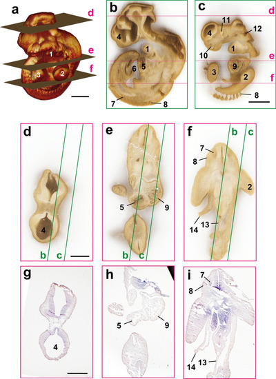
Correlation between 3D image and sections of mouse embryo on E10 in a frozen block. The mouse embryo on E10 was frozen and sliced at a thickness of 10 µm using CoMBI-S system. (a) 3D volume-rendered image using 520 block-face images shows the surface structures of the mouse embryo. (b, c) The reconstructed sagittal planes. (df) Three examples of block-face images are shown. Green lines indicated the positions of (b) and (c). (gi) Sections were collected at the positions of (df). H&E-stained sections can be correlated with block-face images in (df), and the positions where the sections were obtained are indicated as planes in (a). 1: Mandibular arch, 2: Forelimb, 3: Hindlimb, 4: Telencephalon, 5: Right ventricle, 6: Umbilical cord, 7: Dorsal root ganglion, 8: Somite, 9: Left ventricle, 10: Nasal pit, 11: Eye, 12: Otic vesicle, 13: Dorsal mesentery of hindgut, 14: Abdominal wall (broken). Bars: 1 mm (a, c), 500 µm (d, g).

Expression Data

Expression Detail
Antibody Labeling
Phenotype Data

Phenotype Detail
Acknowledgments
This image is the copyrighted work of the attributed author or publisher, and ZFIN has permission only to display this image to its users. Additional permissions should be obtained from the applicable author or publisher of the image. Full text @ Sci. Rep.