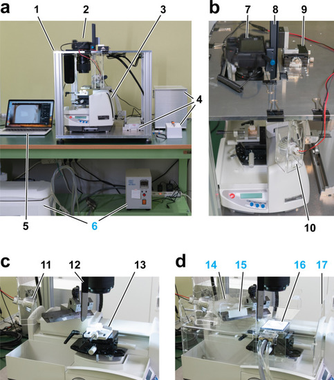
CoMBI system using a sliding microtome. CoMBI-S. (a) The front view of the whole system. The aluminum frame (1) consists of top and bottom plates and four pillars. Camera (2) is attached to the top plate. The sliding type microtome (3) is placed on the bottom plate and its handle is attached to a linear motor (4, left). A microcontroller in the box and switches regulate the linear motor and camera shutter release (4). A laptop computer is used for monitoring the block face and storing images (5). In case of frozen specimen, thermoelectric controller and ice water container are used for cooling a specimen holder (6). (b) The top view of the system. Camera (7) is attached to the top plate via focusing rail for Z positioning (8) and XY positioning stage (9). Two LED lamps at the front and back of specimen illuminate the block face diagonally (10, 11 in c). (c) The side view of the setup for paraffin-embedded specimens. The shading plate (12) and specimen holder (13) are shown. (d) The side view of the setup for frozen specimens. Dry ice for cooling knife (14), frost distributor (15), specimen holder cooling by Peltier element (16), and plastic walls for keeping the cutting environment cool (17) are shown.
|
