FIGURE

Figure 5

ID
ZDB-FIG-210628-49
Publication
Ishii et al., 2021 - Correlative microscopy and block-face imaging (CoMBI) method for both paraffin-embedded and frozen specimens
Other Figures
All Figure Page
Back to All Figure Page
Figure 5

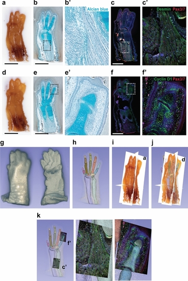
Usability of CoMBI data. Mouse forelimb on E16 were pre-embedded in white-agarose and embedded in paraffin. The 267-serial block-face images with 6-µm interval were obtained. (af) After imaging the 2 block faces shown in (a) and (d), 2 serial sections were collected and used for Alcian blue staining (b, e) or immunostaining (c, f) for desmin (green in c), cyclin D1 (green in f), Pax3/7 (red in c, f). Nuclear DNA was labeled with DAPI (blue in c, f). Boxed areas in (b, c, e, and f) are shown at higher magnification (b′, c′, e′ and f′). (g) Volume-rendered image. (h) Segmentation of cartilages. (i, j) Orthoplanes. Planes labeled with (a) and (d) are identical to block faces shown in (a) and (d). Segmented cartilage and 3D image by volume rendering are also displayed (j). (k) Fluorescent micrographs in (c′) and (f′) are shown in the original location. Bars: 1 mm.

Expression Data

Expression Detail
Antibody Labeling
Phenotype Data

Phenotype Detail
Acknowledgments
This image is the copyrighted work of the attributed author or publisher, and ZFIN has permission only to display this image to its users. Additional permissions should be obtained from the applicable author or publisher of the image. Full text @ Sci. Rep.