|
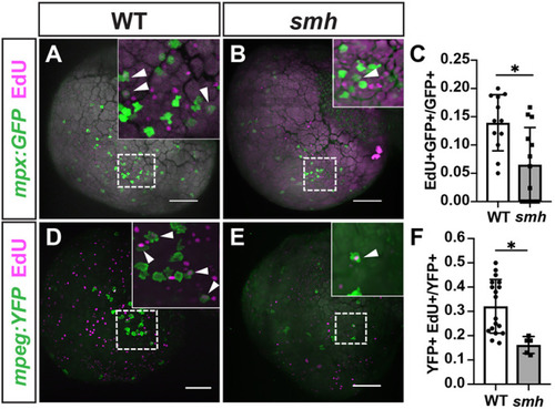
smh myeloid cells are less proliferative. (A,B) Representative images of WT (n=12) and smh (n=19) embryos with mpx:GFP transgene pulsed with EdU at the 20-ss and fixed at 24 hpf. White arrowheads indicate EdU+ cells. (C) Quantification of EdU+/mpx:GFP+ cells from one yolk hemisphere of individual embryos, each data point representing an individual embryo. (D,E) Representative images of WT (n=20) and smh (n=9) embryos with mpeg:YFP transgene pulsed with EdU at the 20-ss and fixed at 24 hpf. White arrowheads indicate EdU+ cells. (F) Quantification of EdU+/mpeg:YFP+ cells from one yolk hemisphere of individual embryos, each data point representing an individual embryo. For C and F, *P<0.05 (two-tailed unpaired Student's t-test). Scale bars: 100 μm.
|