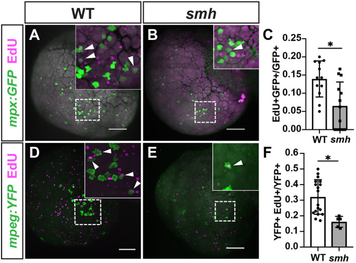
smh myeloid cells are less proliferative. (A,B) Representative images of WT (n=12) and smh (n=19) embryos with mpx:GFP transgene pulsed with EdU at the 20-ss and fixed at 24 hpf. White arrowheads indicate EdU+ cells. (C) Quantification of EdU+/mpx:GFP+ cells from one yolk hemisphere of individual embryos, each data point representing an individual embryo. (D,E) Representative images of WT (n=20) and smh (n=9) embryos with mpeg:YFP transgene pulsed with EdU at the 20-ss and fixed at 24 hpf. White arrowheads indicate EdU+ cells. (F) Quantification of EdU+/mpeg:YFP+ cells from one yolk hemisphere of individual embryos, each data point representing an individual embryo. For C and F, *P<0.05 (two-tailed unpaired Student's t-test). Scale bars: 100 μm.
This image is the copyrighted work of the attributed author or publisher, and
ZFIN has permission only to display this image to its users.
Additional permissions should be obtained from the applicable author or publisher of the image.
Full text @ Dis. Model. Mech.
Your Input Welcome
Thank you for submitting comments. Your input has been emailed to ZFIN curators who may contact you if
additional information is required.
Oops. Something went wrong. Please try again later.