|
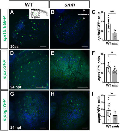
smh mutant embryos have fewer primitive myeloid cells at 24 hpf. (A,B) Representative images of WT sibling (n=15) and smh embryos (n=5) carrying the spi1b:EGFP transgene at the 20-ss. Inset indicates the region of the embryo being imaged. A, anterior; D, dorsal; P, posterior; V, ventral. (C) Quantification of spi1b:EGFP+ myeloid progenitors from one yolk hemisphere of individual embryos. (D,E) Representative images of WT (n=10) and smh (n=10) embryos with mpx:GFP transgene at 24 hpf. (F) Quantification of mpx:GFP+ cells from one yolk hemisphere of individual embryos. (G,H) Representative images of WT sibling (n=19) and smh (n=9) embryos with mpeg1.1:YFP transgene at 24 hpf. (I) Quantification of mpeg1.1:YFP+ myeloid progenitors from one yolk hemisphere of individual embryos. For graphs in C, F and I, each data point represents an individual embryo. **P<0.005, *P<0.05 (two-tailed unpaired Student's t-test). Scale bars: 100 μm.
|