FIGURE

Figure 1

ID
ZDB-FIG-210623-39
Publication
Liu et al., 2021 - In vivo calcium imaging reveals disordered interictal network dynamics in epileptic stxbp1b zebrafish
Other Figures
All Figure Page
Back to All Figure Page
Figure 1

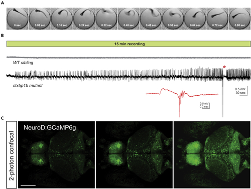
Characterization of epileptic phenotype in stxbp1b mutant zebrafish

(A) Representative spontaneous high-velocity convulsive behavior captured during high-speed imaging (250 fps) of a single stxbp1b mutant larva freely swimming in embryo media at room temperature. Note these were rare events that occur with a velocity near, or greater than, the acquisition speed of the camera (QImaging Optimos cMOS).

(B) Representative 15 min local field potential recordings from randomly selected larvae from a cross of stxbp1b+/- adult breeders. LFP recordings were obtained from a glass microelectrode positioned under a microscope in the midbrain of agarose-embedded larvae at 5 dpf. Larvae were freed from the agarose and genotyped post hoc. Note the presence of small amplitude events building to a large amplitude multi-spike ictal event with postictal depression in the stxbp1b mutant larvae (see inset) but not the WT sibling. These recordings are representative of the spontaneous and unprovoked seizure activity associated with this zebrafish line.

(C) Confocal images taken with a 2-photon microscope of a representative neurod1:GCaMP6f expressing larval zebrafish at three different levels through the central nervous system. Scale bar, 100 μm

Expression Data

Expression Detail
Antibody Labeling
Phenotype Data
Fish:
Observed In:
Stage Range: Day 5 to Days 7-13

Phenotype Detail
Acknowledgments
This image is the copyrighted work of the attributed author or publisher, and ZFIN has permission only to display this image to its users. Additional permissions should be obtained from the applicable author or publisher of the image. Full text @ iScience