|
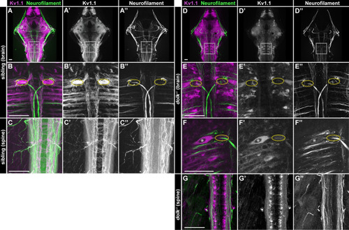
Dolk is required for proper localization of Kv1.1. (A-C) In 5 dpf dolk sibling larvae, Kv1.1 (magenta:A,B,C; grey:A’,B’,C’) localizes to fiber tracts (α-3A10 antibody stains neurofilament; green:A,B,C; grey:A”,B”,C”) throughout the brain (A; box indicates hindbrain zoom in B) and spinal cord (C). Particularly high accumulation of Kv1.1 is observed at the axon cap (B, yellow dotted circle), where spiral fiber neurons form axo-axonic synapses with the Mauthner cell. (D-G) In dolk mutants, Kv1.1 is localized within somata of neurons throughout the brain and spinal cord and is strongly reduced along axons throughout the hindbrain (D,D’,E,E’). Soma localization is particularly apparent in the large Mauthner neuron, and Kv1.1 is strikingly absent from its axon cap (F, nucleus marked by asterisk, E,F axon cap marked by dotted ovals). Scale bars = 50 μM.
|