
Figure 3
- ID
- ZDB-FIG-210617-22
- Publication
- Ellman et al., 2021 - Apex Resection in Zebrafish (Danio rerio) as a Model of Heart Regeneration: A Video-Assisted Guide
- Other Figures
- All Figure Page
- Back to All Figure Page
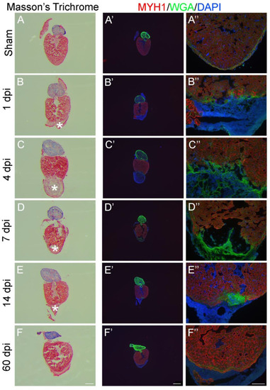
Evaluation of zebrafish heart regeneration in 2D-analysis using histology and immunofluorescence. Cryosections from either sham operated (A,A’,A’’) or apex resected (B–F, B’–F’, B’’–F’’). Zebrafish hearts were stained with Masson’s Trichrome (A–F) or immunostained with anti-MYH1 against myosin heavy chain (MYH1, red), wheat germ agglutinin (WGA, green) and 4′,6′-diamidino-2-phenylindole (DAPI, blue) (A’–F’ and A’’–F’’). Five timepoints (dpi: days post injury) after apex resection were investigated: 1 dpi (B,B’,B’’), 4 dpi (C,C’,C’’), 7 dpi (D,D’,D’’), 14 dpi (E,E’,E’’) and 60 dpi (F,F’,F’’). * marks fibrin clot. Scalebars: A–F = 300 µm, A’–F’ = 500 µm and A’’–F’’ = 100 µm. |