
- Title
-
Apex Resection in Zebrafish (Danio rerio) as a Model of Heart Regeneration: A Video-Assisted Guide
- Authors
- Ellman, D.G., Slaiman, I.M., Mathiesen, S.B., Andersen, K.S., Hofmeister, W., Ober, E.A., Andersen, D.C.
- Source
- Full text @ Int. J. Mol. Sci.
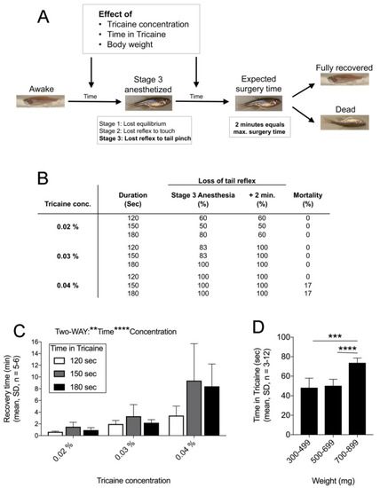
Anaesthesia depends on time and bodyweight of zebrafish. (A) Schematic representation of experimental design. Recovery was tested after the indicated time in Tricaine plus 2 min expected surgery time. (B) Testing the effect of Tricaine concentrations (anaesthesia, rows) and durations (second column) on time to loss of tail reflex—stage 3 (third column) and after (fourth column) +2 min surgery simulation. The zebrafish mortality was noted for each group. (C) Time required to recover after 120 (white bar), 150-(grey bar) or 180 s (black bar) in 0.02, 0.03 or 0.04% Tricaine is depicted, respectively. (D) Time needed to reach stage 3 anaesthesia was measured for each zebrafish weight-class (300–499 mg, 500–699 mg or 700–899 mg). Statistical analysis used include (C) two-way ANOVA and (D) one-way ANOVA with Tukey’s multiple post-test (** p ≤ 0.01, *** p ≤ 0.001, **** p ≤ 0.0001). |
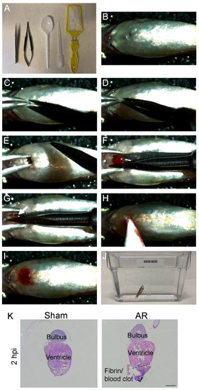
Surgical procedure for sham operation and apex resection (AR). (A) Instruments and utensils required for AR and sham surgery in zebrafish, details are specified in materials and methods. (B–J) Pictures demonstrating each major step in AR surgery (See Supplementary Video S2 for AR and Supplementary Video S3 for sham surgery). (B) Locating the beating heart. (C) Use the forceps to gently lift up the skin. (D) With the micro scissors make a small incision just above the heart. (E) Widen the incision with the forceps. (F) Gently applied pressure on the sides of the incision will make the heart “pop out”. (G) 10–20% of the apex is resected, white arrow marks the resected piece. (H) Remove the instruments and let the heart get back in place before wiping away the blood. (I) The wound after the procedure. (J) The recovering zebrafish (See Supplementary Video S4 for details regarding recovery). (K) Haematoxylin and eosin (HE) stained section from sham and AR hearts 2 h post injury (hpi) showing the heart and where the bulbus should be located as well as the fibrin/blood clot at the resection site. Scalebar: K = 300 µm. |
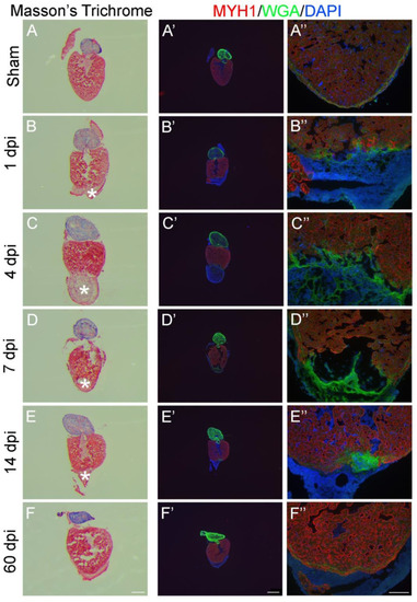
Evaluation of zebrafish heart regeneration in 2D-analysis using histology and immunofluorescence. Cryosections from either sham operated (A,A’,A’’) or apex resected (B–F, B’–F’, B’’–F’’). Zebrafish hearts were stained with Masson’s Trichrome (A–F) or immunostained with anti-MYH1 against myosin heavy chain (MYH1, red), wheat germ agglutinin (WGA, green) and 4′,6′-diamidino-2-phenylindole (DAPI, blue) (A’–F’ and A’’–F’’). Five timepoints (dpi: days post injury) after apex resection were investigated: 1 dpi (B,B’,B’’), 4 dpi (C,C’,C’’), 7 dpi (D,D’,D’’), 14 dpi (E,E’,E’’) and 60 dpi (F,F’,F’’). * marks fibrin clot. Scalebars: A–F = 300 µm, A’–F’ = 500 µm and A’’–F’’ = 100 µm. |
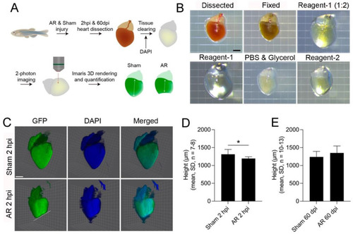
Quantification of heart outgrowth using 3D-analysis of whole zebrafish hearts. (A) Schematic of the experiment design from heart dissection through tissue clearing to Imaris analysis of 2-photon images. (B) Stereomicroscopy of a zebrafish heart during the different stages of tissue clearing with ScaleCUBIC reagent-1 and -2 [29]. Scale bar: 500 µm. (C) 3D images of sham operated or apex resected tg(myl7:EGFP) zebrafish hearts, expressing green fluorescense protein (GFP) under the control of the cardiac myosin light chain 2 promoter, thus marking the myocardium (green) 2 h post injury (hpi). Hearts were co-stained with 4′,6′-diamidino-2-phenylindole (DAPI, blue) and images generated by Imaris. Scale bar: 500 µm. (D,E) Based on the IMARIS analysis, the height of the heart was measured from base to apex in sham operated and apex resected zebrafish hearts, (D) 2 hpi and (E) 60 days post injury (dpi). Statistical analysis included: Normality was tested followed by a Student’s t-test, * p ≤ 0.05. |