
Figure 4
- ID
- ZDB-FIG-210617-23
- Publication
- Ellman et al., 2021 - Apex Resection in Zebrafish (Danio rerio) as a Model of Heart Regeneration: A Video-Assisted Guide
- Other Figures
- All Figure Page
- Back to All Figure Page
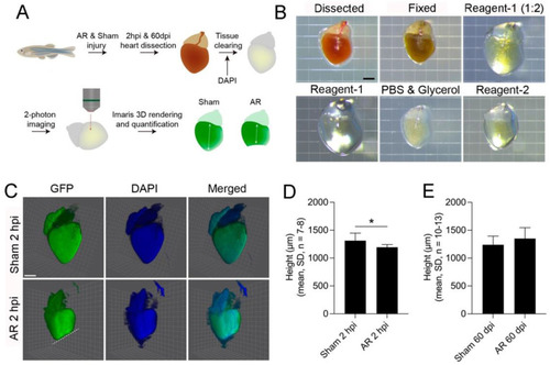
Quantification of heart outgrowth using 3D-analysis of whole zebrafish hearts. (A) Schematic of the experiment design from heart dissection through tissue clearing to Imaris analysis of 2-photon images. (B) Stereomicroscopy of a zebrafish heart during the different stages of tissue clearing with ScaleCUBIC reagent-1 and -2 [29]. Scale bar: 500 µm. (C) 3D images of sham operated or apex resected tg(myl7:EGFP) zebrafish hearts, expressing green fluorescense protein (GFP) under the control of the cardiac myosin light chain 2 promoter, thus marking the myocardium (green) 2 h post injury (hpi). Hearts were co-stained with 4′,6′-diamidino-2-phenylindole (DAPI, blue) and images generated by Imaris. Scale bar: 500 µm. (D,E) Based on the IMARIS analysis, the height of the heart was measured from base to apex in sham operated and apex resected zebrafish hearts, (D) 2 hpi and (E) 60 days post injury (dpi). Statistical analysis included: Normality was tested followed by a Student’s t-test, * p ≤ 0.05. |