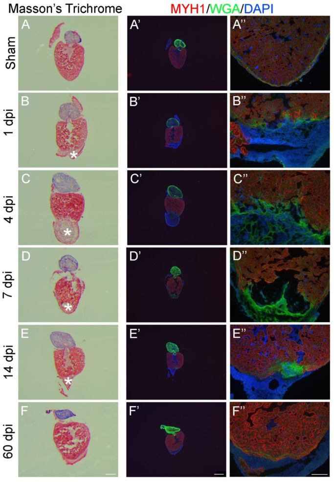
Figure 3 Evaluation of zebrafish heart regeneration in 2D-analysis using histology and immunofluorescence. Cryosections from either sham operated (A,A’,A’’) or apex resected (B–F, B’–F’, B’’–F’’). Zebrafish hearts were stained with Masson’s Trichrome (A–F) or immunostained with anti-MYH1 against myosin heavy chain (MYH1, red), wheat germ agglutinin (WGA, green) and 4′,6′-diamidino-2-phenylindole (DAPI, blue) (A’–F’ and A’’–F’’). Five timepoints (dpi: days post injury) after apex resection were investigated: 1 dpi (B,B’,B’’), 4 dpi (C,C’,C’’), 7 dpi (D,D’,D’’), 14 dpi (E,E’,E’’) and 60 dpi (F,F’,F’’). * marks fibrin clot. Scalebars: A–F = 300 µm, A’–F’ = 500 µm and A’’–F’’ = 100 µm.
                
                    
                        Image


                        Figure Caption


                        Acknowledgments


This image is the copyrighted work of the attributed author or publisher, and
ZFIN has permission only to display this image to its users.
Additional permissions should be obtained from the applicable author or publisher of the image.
      Full text @ Int. J. Mol. Sci.