
Fig. 5
- ID
- ZDB-FIG-210520-56
- Publication
- Mitchell et al., 2021 - The alx3 gene shapes the zebrafish neurocranium by regulating frontonasal neural crest cell differentiation timing
- Other Figures
- All Figure Page
- Back to All Figure Page
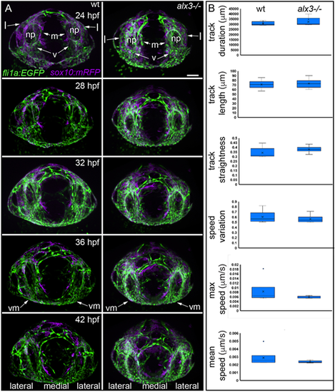
(A) Still frames from time-lapse recordings of craniofacial progenitors. At 24 hpf, NCCs that are lateral (l), medial (m) and ventral (v) to the nasal placode (np) express alx3 (see Fig. 2C-E). Arrows indicate that postmigratory NCCs are present in both wild types (wt) and mutants. At 36 hpf, NCCs ventral and medial (vm) to the eye express alx3 (see Fig. 3B). Arrows illustrate that these cells are present in both wild types and alx3 mutants. All images are frontal view. (B) Semi-automated quantification of cell movement reveals no significant differences (unpaired t-test) between wild types and mutants. We tracked multiple cell movement parameters from 728 wild-type and 886 alx3 mutant cells from six embryos of each genotype. For box plots, boxes represent the interquartile range, whiskers are the data range, the lines are the medians and x represents the mean. Scale bar: 60 µm. |