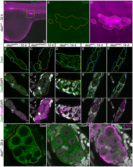
(A-A″) Dazl is not detectable in PGCs marked with Vasa at 30 hpf. (A′,A″) Enlargement of the yellow dotted box in A with Dazl (A′, green) and Vasa (A″, magenta). (B-F″) Single confocal plane of representative 12-day dazlae57/+ and dazlae57/ae57 gonads (B-C″), and dazl+/+, dazlΔ7/+ or dazlΔ7/Δ7 14-day gonads (D-F″) immunostained with Dazl (green), Vasa (magenta) and DAPI (gray) as nuclear marker. Yellow dotted lines delineate GCs and blue lines outline somatic cells. (B-C″) Localization of Dazl in Vasa+ GC of dazlae57/+ (B-B″, n=1) and dazlae57/ae57 (C-C″, n=1). There was less abundant Dazl in dazlae57/ae57 gonads. (D-D″) Localization of Dazl in Vasa+ GC of dazl+/+ (n=1). Image is 63× without 2.5× zoom to show full gonad at resolution 512×512. (E-E″) Localization of Dazl in Vasa+ GC of dazlΔ7/+ (n=1). (F-F″) No detecatable Dazl in Vasa+ GC of dazlΔ7/Δ7 (n=2). Yellow dotted line delineates the gonad. (G-G″) Twenty-nine-day gonads labeled with Dazl (green) and Vasa (magenta). DAPI (gray) marks nuclei. Dazl protein was diffusely localized in the cytoplasm of the GCs.
|
