
Fig. 4
- ID
- ZDB-FIG-210504-19
- Publication
- Peng et al., 2020 - Induction of Wnt signaling antagonists and p21-activated kinase enhances cardiomyocyte proliferation during zebrafish heart regeneration
- Other Figures
- All Figure Page
- Back to All Figure Page
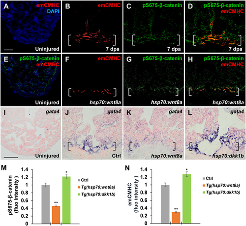
Wnt signaling inhibition associates with CM dedifferentiation during heart regeneration. (A‒H) Immunofluorescence analyses showing increased emCMHC stained with N2.261 antibody and pS675-β-catenin at apical myocardial cells of the wound at 7 dpa (A‒D), which was reduced by wnt8a activation in injured Tg(hsp70:wnt8a) hearts (E‒H). N2.261 is not detectable in uninjured hearts (A and E) and overlaps mostly with induced pS675-β-catenin (D). (D and H) Merged panels of B and C and F and G, respectively. (I‒L) ISH analyses displaying gata4 expression at injured myocardial cell edges in heat-shocked Ctrl hearts (J) at 7 dpa, which was reduced in Tg(hsp:wnt8a) hearts (K) and increased in Tg(hsp:dkk1b) hearts (L) but hardly detectable in uninjured hearts (I). Brackets indicate amputation planes. Scale bar, 100 µm. (M and N) Bar charts depicting pS675-β-catenin (M) and emCMHC (N) levels in Ctrl (normalized as 1), Tg(hsp:wnt8a), and Tg(hsp:dkk1b) hearts. Fluorescent intensities were measured at the injury border zone using Image J. Data are mean ± SEM from five hearts for each group. Student’s t-test, *P < 0.05, **P < 0.01. |