
Fig. 3
- ID
- ZDB-FIG-210504-18
- Publication
- Peng et al., 2020 - Induction of Wnt signaling antagonists and p21-activated kinase enhances cardiomyocyte proliferation during zebrafish heart regeneration
- Other Figures
- All Figure Page
- Back to All Figure Page
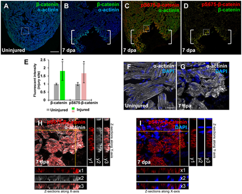
Induction of pS675-β-catenin at disassembled sarcomeres in the injured myocardium following cardiac damage. (A) In uninjured hearts, β-catenin is detectable throughout the myocardium stained with a sarcomeric Z-disk marker α-actinin. (B‒D) Following ventricular resection, β-catenin (B) and pS675-β-catenin (C) are induced at the apical cell edge of wounded myocardia. (D) Merged panel of B and C without α-actinin. Brackets, amputation area. (E) Bar chart depicting β-catenin and pS675-β-catenin levels following ventricular resection. Fluorescent intensities were measured at the injury border zone using Image J. β-catenin or pS675-β-catenin levels in control hearts are normalized as 1. Data are mean ± SEM from five hearts for each group. Student’s t-test, *P < 0.05. (F) High-magnification image of the dash-lined area in A displaying CMs in organized sarcomeric arrays in uninjured hearts. (G‒I) High-magnification images of the dash-lined window in C exhibiting the co-localization of pS675-β-catenin (H and I) with disassembled sarcomeres (G and H) in the injured myocardial cell edge. (I) Merged panel of G and H without α-actinin. 3D analyses display co-localizations of pS675-β-catenin (red) with dissociated sarcomere components (white) along the X-axis (H, x1‒x3) and the Y-axis (H, y1‒y3), as well as the cytoplasmic localization of pS675-β-catenin in Z sections (I, x1‒x3 and y1‒y3). Scale bar, 100 µm (A‒D) and 10 µm (F‒I). |