FIGURE

Fig. 3

ID
ZDB-FIG-210419-3
Publication
Don et al., 2021 - In vivo Validation of Bimolecular Fluorescence Complementation (BiFC) to Investigate Aggregate Formation in Amyotrophic Lateral Sclerosis (ALS)
Other Figures
All Figure Page
Back to All Figure Page
Fig. 3

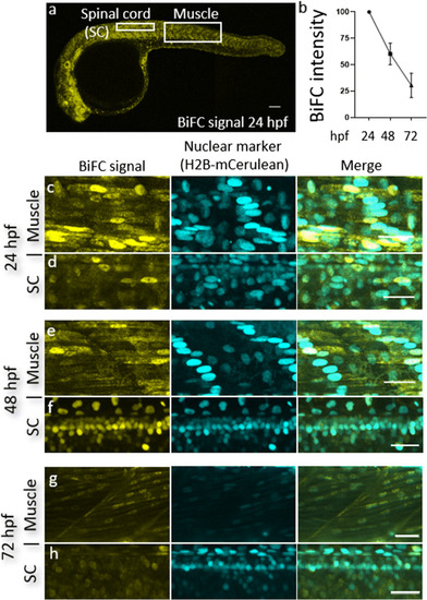
TDP-43 BiFC can be detected in muscle cells and motor neurons and is predominantly nuclear. a A representative image of a 24 hpf embryo injected with our TDP-43 BiFC constructs. Scale bar, 200 μm. b Fluorescent intensity as a percentage of signal at 24 hpf. c, d Representative pictures of the human wild-type TDP-43 BiFC signal at 24 hpf in muscle cells (c) and spinal cord motor neurons (d). e, f Representative pictures of the human wild-type TDP-43 BiFC signal at 48 hpf in muscle cells (e) and spinal cord motor neurons (f). g, h Representative pictures of the human wild-type TDP-43 BiFC signal at 72 hpf in muscle cells (g) and spinal cord motor neurons (h). Scale bar, 30 μm

Expression Data

Expression Detail
Antibody Labeling
Phenotype Data

Phenotype Detail
Acknowledgments
This image is the copyrighted work of the attributed author or publisher, and ZFIN has permission only to display this image to its users. Additional permissions should be obtained from the applicable author or publisher of the image. Full text @ Mol. Neurobiol.