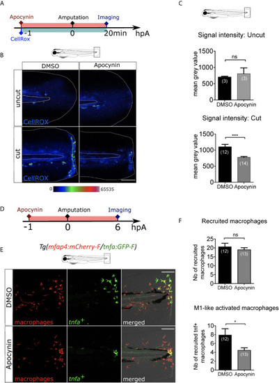
ROS release at the wound mediate macrophage activation but not recruitment. (A) Schedule of the experiment. 1h before the fin fold injury at 3 dpf, larvae were incubated in Apocynin or DMSO, containing CellROX solution for the detection of the ROS production. Drug and staining were both removed at 20 min pA, and larvae were immediately imaged using epi-fluorescent microscopy. (B) Representative images of the CellROX fluorescence in uncut or cut fin folds, after the treatment with DMSO or Apocynin at 20 min pA. Rainbow color scale was applied to images, emphasizing the differences in signal intensity. The white lines outline the fin fold and the notochord. Scale bar: 100 μm. (C) Quantification of signal intensity of the CellROX fluorescence by mean gray value. Representative experiment of two independent experiments, mean ± SEM, nlarvae is indicated in brackets, upper graph: Mann Whitney test, two-tailed, ns – not significant, bottom graph: one-tailed t-test with Welch’s correction, ***p<0.001. (D) Schedule of the experiment. From 1 h before the fin fold amputation at 3 dpf, until 6 hpA, Tg(mfap4:mCherry-F/tnfa:GFP-F) larvae were incubated in Apocynin or DMSO, and then imaged at 6 hpA using confocal microscopy. (E) Tail images are representative maximum projections of the fluorescence of mCherry-F (macrophages), GFP-F (tnfa+ cells) and merged channel images with brightfield after the treatment with DMSO (up) or Apocynin (down) at 6 hpA. Scale bars: 100 μm. (F) Quantification of recruited macrophages (up) and tnfa+ recruited macrophages (down) after DMSO and Apocynin treatments at 6 hpA. Representative experiment of two independent experiments, mean ± SEM, nlarvae is indicated in brackets, one-tailed t-test with Welch’s correction, *p<0.05.
|
